Structural insights into the octamerization of glycerol dehydrogenase.
Park, T., Kang, J.Y., Jin, M., Yang, J., Kim, H., Noh, C., Jung, C.H., Eom, S.H.(2024) PLoS One 19: e0300541-e0300541
- PubMed: 38483875 Search on PubMedSearch on PubMed Central
- DOI: https://doi.org/10.1371/journal.pone.0300541
- Primary Citation Related Structures:
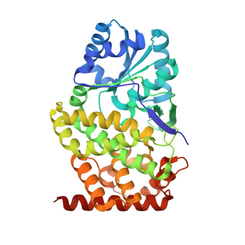
8X6M - PubMed Abstract:
Glycerol dehydrogenase (GDH) catalyzes glycerol oxidation to dihydroxyacetone in a NAD+-dependent manner. As an initiator of the oxidative pathway of glycerol metabolism, a variety of functional and structural studies of GDH have been conducted previously. Structural studies revealed intriguing features of GDH, like the flexible β-hairpin and its significance. Another commonly reported structural feature is the enzyme's octameric oligomerization, though its structural details and functional significance remained unclear. Here, with a newly reported GDH structure, complexed with both NAD+ and glycerol, we analyzed the octamerization of GDH. Structural analyses revealed that octamerization reduces the structural dynamics of the N-domain, which contributes to more consistently maintaining a distance required for catalysis between the cofactor and substrate. This suggests that octamerization may play a key role in increasing the likelihood of the enzyme reaction by maintaining the ligands in an appropriate configuration for catalysis. These findings expand our understanding of the structure of GDH and its relation to the enzyme's activity.
- Department of Chemistry, Gwangju Institute of Science and Technology (GIST), Gwangju, Republic of Korea.
Organizational Affiliation: