8WXB

Cryo-EM structure of the alpha-carboxysome shell vertex from Prochlorococcus MED4


Experimental Data Snapshot

  • Method: ELECTRON MICROSCOPY
  • Resolution: 4.20 Å
  • Aggregation State: PARTICLE 
  • Reconstruction Method: SINGLE PARTICLE 

wwPDB Validation   3D Report Full Report


This is version 1.1 of the entry. See complete history


Literature

Structure and assembly of the alpha-carboxysome in the marine cyanobacterium Prochlorococcus.

Zhou, R.Q.Jiang, Y.L.Li, H.Hou, P.Kong, W.W.Deng, J.X.Chen, Y.Zhou, C.Z.Zeng, Q.

(2024) Nat Plants 10: 661-672

  • DOI: https://doi.org/10.1038/s41477-024-01660-9
  • Primary Citation of Related Structures:  
    8WXB

  • PubMed Abstract: 

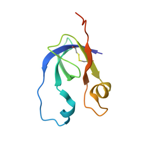
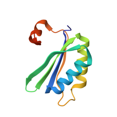
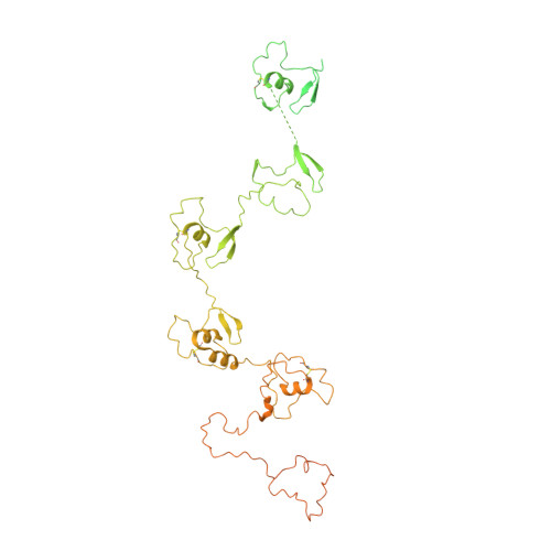
    Carboxysomes are bacterial microcompartments that encapsulate the enzymes RuBisCO and carbonic anhydrase in a proteinaceous shell to enhance the efficiency of photosynthetic carbon fixation. The self-assembly principles of the intact carboxysome remain elusive. Here we purified α-carboxysomes from Prochlorococcus and examined their intact structures using single-particle cryo-electron microscopy to solve the basic principles of their shell construction and internal RuBisCO organization. The 4.2 Å icosahedral-like shell structure reveals 24 CsoS1 hexamers on each facet and one CsoS4A pentamer at each vertex. RuBisCOs are organized into three concentric layers within the shell, consisting of 72, 32 and up to 4 RuBisCOs at the outer, middle and inner layers, respectively. We uniquely show how full-length and shorter forms of the scaffolding protein CsoS2 bind to the inner surface of the shell via repetitive motifs in the middle and C-terminal regions. Combined with previous reports, we propose a concomitant 'outside-in' assembly principle of α-carboxysomes: the inner surface of the self-assembled shell is reinforced by the middle and C-terminal motifs of the scaffolding protein, while the free N-terminal motifs cluster to recruit RuBisCO in concentric, three-layered spherical arrangements. These new insights into the coordinated assembly of α-carboxysomes may guide the rational design and repurposing of carboxysome structures for improving plant photosynthetic efficiency.


  • Organizational Affiliation

    Department of Ocean Science, The Hong Kong University of Science and Technology, Clear Water Bay, Hong Kong, China.


Macromolecules
Find similar proteins by:  (by identity cutoff)  |  3D Structure
Entity ID: 1
MoleculeChains Sequence LengthOrganismDetailsImage
Carboxysome shell vertex protein CsoS4AA [auth 2]88Prochlorococcus marinus subsp. pastoris str. CCMP1986Mutation(s): 0 
UniProt
Find proteins for Q7V2C6 (Prochlorococcus marinus subsp. pastoris (strain CCMP1986 / NIES-2087 / MED4))
Explore Q7V2C6 
Go to UniProtKB:  Q7V2C6
Entity Groups  
Sequence Clusters30% Identity50% Identity70% Identity90% Identity95% Identity100% Identity
UniProt GroupQ7V2C6
Sequence Annotations
Expand
  • Reference Sequence
Find similar proteins by:  (by identity cutoff)  |  3D Structure
Entity ID: 2
MoleculeChains Sequence LengthOrganismDetailsImage
Major carboxysome shell protein CsoS198Prochlorococcus marinus subsp. pastoris str. CCMP1986Mutation(s): 0 
UniProt
Find proteins for Q7V2D1 (Prochlorococcus marinus subsp. pastoris (strain CCMP1986 / NIES-2087 / MED4))
Explore Q7V2D1 
Go to UniProtKB:  Q7V2D1
Entity Groups  
Sequence Clusters30% Identity50% Identity70% Identity90% Identity95% Identity100% Identity
UniProt GroupQ7V2D1
Sequence Annotations
Expand
  • Reference Sequence
Find similar proteins by:  (by identity cutoff)  |  3D Structure
Entity ID: 3
MoleculeChains Sequence LengthOrganismDetailsImage
Carboxysome assembly protein CsoS2AA [auth Z],
Z [auth Y]
765Prochlorococcus marinus subsp. pastoris str. CCMP1986Mutation(s): 0 
UniProt
Find proteins for Q7V2C8 (Prochlorococcus marinus subsp. pastoris (strain CCMP1986 / NIES-2087 / MED4))
Explore Q7V2C8 
Go to UniProtKB:  Q7V2C8
Entity Groups  
Sequence Clusters30% Identity50% Identity70% Identity90% Identity95% Identity100% Identity
UniProt GroupQ7V2C8
Sequence Annotations
Expand
  • Reference Sequence
Experimental Data & Validation

Experimental Data

  • Method: ELECTRON MICROSCOPY
  • Resolution: 4.20 Å
  • Aggregation State: PARTICLE 
  • Reconstruction Method: SINGLE PARTICLE 

Structure Validation

View Full Validation Report


Entry History & Funding Information

Deposition Data


Funding OrganizationLocationGrant Number
Chinese Academy of SciencesChinaXDA24020302
Chinese Academy of SciencesChinaXDB37020301
The University Grants Committee, Research Grants Council (RGC)Hong KongHKUST C6012-22GF
National Natural Science Foundation of China (NSFC)China32241025
National Natural Science Foundation of China (NSFC)China32171198

Revision History  (Full details and data files)

  • Version 1.0: 2024-01-31
    Type: Initial release
  • Version 1.1: 2024-05-22
    Changes: Database references