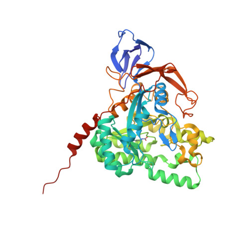
The complexed crystal structure of dihydropyrimidinase reveals a potential interactive link with the neurotransmitter gamma-aminobutyric acid (GABA).
Huang, Y.H., Huang, C.Y.(2024) Biochem Biophys Res Commun 692: 149351-149351
- PubMed: 38056157 Search on PubMed
- DOI: https://doi.org/10.1016/j.bbrc.2023.149351
- Primary Citation Related Structures:
8WQ9 - PubMed Abstract:
Dihydropyrimidinase (DHPase) plays a crucial role in pyrimidine degradation, showcasing a broad substrate specificity that extends beyond pyrimidine catabolism, hinting at additional roles for this ancient enzyme. In this study, we solved the crystal structure of Pseudomonas aeruginosa DHPase (PaDHPase) complexed with the neurotransmitter γ-aminobutyric acid (GABA) at a resolution of 1.97 Å (PDB ID 8WQ9). Our structural analysis revealed two GABA binding sites in each monomer of PaDHPase. Interactions between PaDHPase and GABA molecules, involving residues within a contact distance of <4 Å, were examined. In silico analyses via PISA and PLIP software revealed hydrogen bonds formed between the side chain of Cys318 and GABA 1, as well as the main chains of Ser333, Ile335, and Asn337 with GABA 2. Comparative structural analysis between GABA-bound and unbound states unveiled significant conformational changes at the active site, particularly within dynamic loop I, supporting the conclusion that PaDHPase binds GABA through the loop-out mechanism. Building upon this molecular evidence, we discuss and propose a working model. The study expands the GABA interactome by identifying DHPase as a novel GABA-interacting protein and provides structural insight into the interaction between a dimetal center in the protein's active site and GABA. Further investigations are warranted to explore potential interactions of GABA with other DHPase-like proteins and to understand whether DHPase may have additional regulatory and physiological roles in the cell, extending beyond pyrimidine catabolism.
- Department of Biomedical Sciences, Chung Shan Medical University, No.110, Sec.1, Chien-Kuo N. Rd., Taichung City, Taiwan.
Organizational Affiliation: