8WDT | pdb_00008wdt

Crystal structure of the human adenosine A2A receptor in complex with photoresponsive ligand photoNECA(blue)


Experimental Data Snapshot

  • Method: X-RAY DIFFRACTION
  • Resolution: 3.34 Å
  • R-Value Free: 
    0.266 (Depositor), 0.265 (DCC) 
  • R-Value Work: 
    0.214 (Depositor), 0.214 (DCC) 
  • R-Value Observed: 
    0.219 (Depositor) 

Starting Models: experimental
View more details

wwPDB Validation 3D Report Full Report

Validation slider image for 8WDT

Ligand Structure Quality Assessment 


This is version 1.1 of the entry. See complete history

Literature

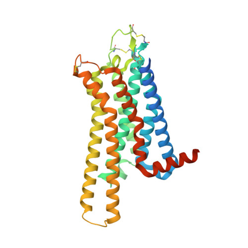
Crystal structure reveals the binding mode and selectivity of a photoswitchable ligand for the adenosine A 2A receptor.

Araya, T.Matsuba, Y.Suzuki, H.Doura, T.Nuemket, N.Nango, E.Yamamoto, M.Im, D.Asada, H.Kiyonaka, S.Iwata, S.

(2023) Biochem Biophys Res Commun 695: 149393-149393

  • DOI: https://doi.org/10.1016/j.bbrc.2023.149393
  • Primary Citation Related Structures: 
    8WDT

  • PubMed Abstract: 

    Rational synthetic expansion of photoresponsive ligands is important for photopharmacological studies. Adenosine A 2A receptor (A 2A R) is stimulated by adenosine and related in Parkinson's disease and other diseases. Here, we report the crystal structure of the A 2A R in complex with the novel photoresponsive ligand photoNECA (blue) at 3.34 Å resolution. PhotoNECA (blue) was designed for this structural study and the cell-based assay showed a photoresponsive and receptor selective characteristics of photoNECA (blue) for A 2A R. The crystal structure explains the binding mode, photoresponsive mechanism and receptor selectivity of photoNECA (blue). Our study would promote not only the rational design of photoresponsive ligands but also dynamic structural studies of A 2A R.


  • Organizational Affiliation
    • Department of Cell Biology, Graduate School of Medicine, Kyoto University, Kyoto, 606-8501, Japan.

Macromolecule Content 

  • Total Structure Weight: 87.2 kDa 
  • Atom Count: 5,667 
  • Modeled Residue Count: 734 
  • Deposited Residue Count: 788 
  • Unique protein chains: 3

Macromolecules

Find similar proteins by:|  3D Structure
Entity ID: 1
MoleculeChains  Sequence LengthOrganismDetailsImage
Adenosine receptor A2a351Homo sapiensMutation(s): 6 
Gene Names: ADORA2A
Membrane Entity: Yes 
UniProt & NIH Common Fund Data Resources
Find proteins for P29274 (Homo sapiens)
Explore P29274 
Go to UniProtKB:  P29274
PHAROS:  P29274
GTEx:  ENSG00000128271 
Entity Groups
Sequence Clusters30% Identity50% Identity70% Identity90% Identity95% Identity100% Identity
UniProt GroupP29274
Sequence Annotations
Expand
Reference Sequence
Find similar proteins by:|  3D Structure
Entity ID: 2
MoleculeChains  Sequence LengthOrganismDetailsImage
Antibody Fab fragment light chain213Mus musculusMutation(s): 0 
Find similar proteins by:|  3D Structure
Entity ID: 3
MoleculeChains  Sequence LengthOrganismDetailsImage
Antibody Fab fragment heavy chain224Mus musculusMutation(s): 0 

Small Molecules

Ligands 1 Unique
IDChains Name / Formula / InChI Key2D Diagram3D Interactions
WCH
(Subject of Investigation/LOI)

Query on WCH


Download:Ideal Coordinates CCD File
D [auth A](2S,3S,4R,5R)-5-(6-amino-2-((E)-phenyldiazenyl)-9H-purin-9-yl)-N-ethyl-3,4-dihydroxytetrahydrofuran-2-carboxamide
C18 H20 N8 O4
TWQRPAGBMNXURX-APJVYBOUSA-N

Experimental Data & Validation

Experimental Data

  • Method: X-RAY DIFFRACTION
  • Resolution: 3.34 Å
  • R-Value Free:  0.266 (Depositor), 0.265 (DCC) 
  • R-Value Work:  0.214 (Depositor), 0.214 (DCC) 
  • R-Value Observed: 0.219 (Depositor) 
Space Group: C 1 2 1
Unit Cell:
Length ( Å )Angle ( ˚ )
a = 372.64α = 90
b = 52.52β = 94.79
c = 63.4γ = 90
Software Package:
Software NamePurpose
PHENIXrefinement
XDSdata reduction
XSCALEdata scaling
PHENIXphasing
Cootmodel building
PDB_EXTRACTdata extraction

Structure Validation

View Full Validation Report


Ligand Structure Quality Assessment 


Entry History 

& Funding Information

Deposition Data


Funding OrganizationLocationGrant Number
Japan Society for the Promotion of Science (JSPS)Japan--
Japan Agency for Medical Research and Development (AMED)Japan--

Revision History  (Full details and data files)

  • Version 1.0: 2024-01-17
    Type: Initial release
  • Version 1.1: 2024-11-13
    Changes: Structure summary