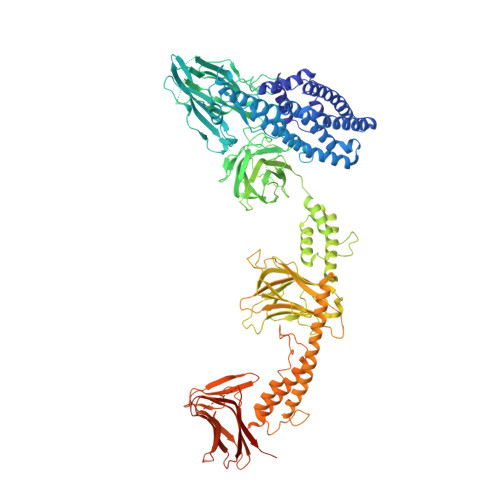
Crystal structure of the in-cell Cry1Aa purified from Bacillus thuringiensis.
Tanaka, J., Abe, S., Hayakawa, T., Kojima, M., Yamashita, K., Hirata, K., Ueno, T.(2023) Biochem Biophys Res Commun 685: 149144-149144
- PubMed: 37922785 Search on PubMed
- DOI: https://doi.org/10.1016/j.bbrc.2023.149144
- Primary Citation Related Structures:
8W7N - PubMed Abstract:
In-cell protein crystals which spontaneously crystallize in living cells, have recently been analyzed in investigations of their structures and biological functions. The crystals have been challenging to analyze structurally because of their small size. Therefore, the number of in-cell protein crystals in which the native structure has been determined is limited because most of the structures of in-cell crystals have been determined by recrystallization after dissolution. Some proteins have been reported to form intermolecular disulfide bonds in natural protein crystals that stabilize the crystals. Here, we focus on Cry1Aa, a cysteine-rich protein that crystallizes in Bacillus thuringiensis (Bt) and forms disulfide bonds. Previously, the full-length structure of 135 kDa Cry1Ac, which is the same size as Cry1Aa, was determined by recrystallization of dissolved protein from crystals purified from Bt cells. However, the formation of disulfide bonds has not been investigated because it was necessary to replace cysteine residues to prevent aggregation of the soluble protein. In this work, we succeeded in direct X-ray crystallographic analysis using crystals purified from Bt cells and characterized the cross-linked network of disulfide bonds within Cry1Aa crystals.
- School of Life Science and Technology, Tokyo Institute of Technology, Nagatsuta-cho 4259, Midori-ku, Yokohama, 226-8501, Japan.
Organizational Affiliation: