8VZ9

Crystal structure of mouse MAIT M2A TCR-MR1-5-OP-RU complex


Experimental Data Snapshot

  • Method: X-RAY DIFFRACTION
  • Resolution: 3.40 Å
  • R-Value Free: 0.263 
  • R-Value Work: 0.242 
  • R-Value Observed: 0.244 

wwPDB Validation   3D Report Full Report


Ligand Structure Quality Assessment 


This is version 1.0 of the entry. See complete history


Literature

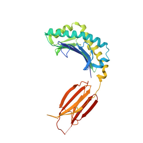
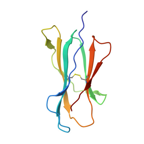
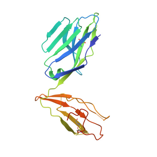
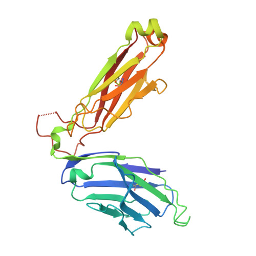
Mouse mucosal-associated invariant T cell receptor recognition of MR1 presenting the vitamin B metabolite, 5-(2-oxopropylideneamino)-6-D-ribitylaminouracil.

Ciacchi, L.Mak, J.Y.W.Le, J.P.Fairlie, D.P.McCluskey, J.Corbett, A.J.Rossjohn, J.Awad, W.

(2024) J Biol Chem : 107229-107229

  • DOI: https://doi.org/10.1016/j.jbc.2024.107229
  • Primary Citation of Related Structures:  
    8VZ8, 8VZ9

  • PubMed Abstract: 

    Mucosal-associated invariant T (MAIT) cells can elicit immune responses against riboflavin-based antigens presented by the evolutionary conserved MHC Class I related-1 protein, MR1. While we have an understanding of the structural basis of human MAIT cell receptor (TCR) recognition of human MR1 presenting a variety of ligands, how the semi-invariant mouse MAIT TCR binds mouse MR1-ligand remains unknown. Here we determine the crystal structures of two mouse TRAV1-TRBV13-2 + MAIT TCR-MR1-5-OP-RU ternary complexes, whose TCRs differ only in the composition of their CDR3β loops. These mouse MAIT TCRs mediate high affinity interactions with mouse MR1-5-OP-RU and cross-recognise human MR1-5-OP-RU. Similarly, a human MAIT TCR could bind mouse MR1-5-OP-RU with high affinity. This cross-species recognition indicates the evolutionary conserved nature of this MAIT TCR-MR1 axis. Comparing crystal structures of the mouse versus human MAIT TCR-MR1-5-OP-RU complexes provides structural insight into the conserved nature of this MAIT TCR-MR1 interaction, and conserved specificity for the microbial antigens, whereby key germline-encoded interactions required for MAIT activation are maintained. This is an important consideration for the development of MAIT cell-based therapeutics that will rely on preclinical mouse models of disease.


  • Organizational Affiliation

    Infection and Immunity Program, Department of Biochemistry and Molecular Biology, Biomedicine Discovery Institute, Monash University, Clayton, Victoria 3800, Australia.


Macromolecules
Find similar proteins by:  (by identity cutoff)  |  3D Structure
Entity ID: 1
MoleculeChains Sequence LengthOrganismDetailsImage
Major histocompatibility complex class I-related gene protein271Mus musculusMutation(s): 0 
Gene Names: Mr1Mr1a
UniProt & NIH Common Fund Data Resources
Find proteins for Q8HWB0 (Mus musculus)
Explore Q8HWB0 
Go to UniProtKB:  Q8HWB0
IMPC:  MGI:1195463
Entity Groups  
Sequence Clusters30% Identity50% Identity70% Identity90% Identity95% Identity100% Identity
UniProt GroupQ8HWB0
Sequence Annotations
Expand
  • Reference Sequence
Find similar proteins by:  (by identity cutoff)  |  3D Structure
Entity ID: 2
MoleculeChains Sequence LengthOrganismDetailsImage
Beta-2-microglobulin100Mus musculusMutation(s): 0 
Gene Names: B2m
UniProt & NIH Common Fund Data Resources
Find proteins for P01887 (Mus musculus)
Explore P01887 
Go to UniProtKB:  P01887
IMPC:  MGI:88127
Entity Groups  
Sequence Clusters30% Identity50% Identity70% Identity90% Identity95% Identity100% Identity
UniProt GroupP01887
Sequence Annotations
Expand
  • Reference Sequence
Find similar proteins by:  (by identity cutoff)  |  3D Structure
Entity ID: 3
MoleculeChains Sequence LengthOrganismDetailsImage
Mouse MAIT TRAV1-TRAJ33 a-chain204Mus musculusMutation(s): 0 
Entity Groups  
Sequence Clusters30% Identity50% Identity70% Identity90% Identity95% Identity100% Identity
Sequence Annotations
Expand
  • Reference Sequence
Find similar proteins by:  (by identity cutoff)  |  3D Structure
Entity ID: 4
MoleculeChains Sequence LengthOrganismDetailsImage
Mouse MAIT MBV13-2A b-chain246Mus musculusMutation(s): 0 
Entity Groups  
Sequence Clusters30% Identity50% Identity70% Identity90% Identity95% Identity100% Identity
Sequence Annotations
Expand
  • Reference Sequence
Small Molecules
Ligands 2 Unique
IDChains Name / Formula / InChI Key2D Diagram3D Interactions
2LJ (Subject of Investigation/LOI)
Query on 2LJ

Download Ideal Coordinates CCD File 
G [auth A]1-deoxy-1-({2,6-dioxo-5-[(E)-propylideneamino]-1,2,3,6-tetrahydropyrimidin-4-yl}amino)-D-ribitol
C12 H20 N4 O6
YCMPUNANLDFPQG-FHZGFTDOSA-N
GOL
Query on GOL

Download Ideal Coordinates CCD File 
E [auth A],
F [auth A]
GLYCEROL
C3 H8 O3
PEDCQBHIVMGVHV-UHFFFAOYSA-N
Experimental Data & Validation

Experimental Data

  • Method: X-RAY DIFFRACTION
  • Resolution: 3.40 Å
  • R-Value Free: 0.263 
  • R-Value Work: 0.242 
  • R-Value Observed: 0.244 
  • Space Group: P 2 21 21
Unit Cell:
Length ( Å )Angle ( ˚ )
a = 64.531α = 90
b = 84.259β = 90
c = 177.584γ = 90
Software Package:
Software NamePurpose
PHENIXrefinement
pointlessdata scaling
XDSdata reduction
PHENIXphasing

Structure Validation

View Full Validation Report


Ligand Structure Quality Assessment 


Entry History & Funding Information

Deposition Data


Funding OrganizationLocationGrant Number
Australian Research Council (ARC)AustraliaDE220101491
Australian Research Council (ARC)AustraliaDP220102401

Revision History  (Full details and data files)

  • Version 1.0: 2024-04-10
    Type: Initial release