8VXJ | pdb_00008vxj

The crystal structure of human apolipoprotein A-I in complex with Fab 55201


Experimental Data Snapshot

  • Method: X-RAY DIFFRACTION
  • Resolution: 2.70 Å
  • R-Value Free: 
    0.298 (Depositor), 0.294 (DCC) 
  • R-Value Work: 
    0.266 (Depositor), 0.262 (DCC) 
  • R-Value Observed: 
    0.268 (Depositor) 

Starting Models: experimental
View more details

wwPDB Validation 3D Report Full Report

Validation slider image for 8VXJ

This is version 1.1 of the entry. See complete history

Literature

The Structure of the Apolipoprotein A-I Monomer Provides Insights Into Its Oligomerisation and Lipid-binding Mechanisms.

Tou, H.I.Rosenes, Z.Khandokar, Y.Zlatic, C.O.Metcalfe, R.D.Mok, Y.F.Morton, C.J.Gooley, P.R.Griffin, M.D.W.

(2025) J Mol Biology 437: 169394-169394

  • DOI: https://doi.org/10.1016/j.jmb.2025.169394
  • Primary Citation Related Structures: 
    8VXJ

  • PubMed Abstract: 

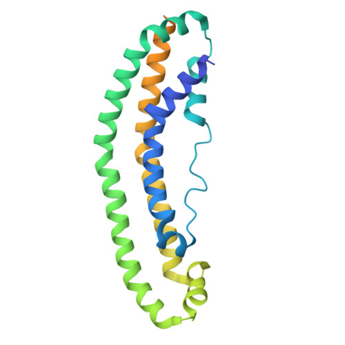
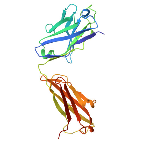
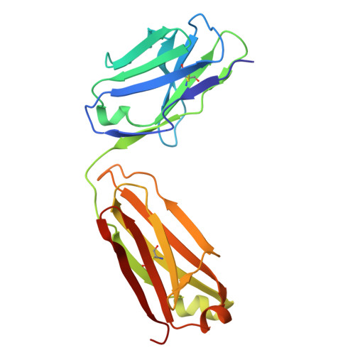
    Apolipoprotein A-I (apoA-I) plays important roles in clearing cholesterol and phospholipids from peripheral tissues, forming high-density lipoprotein (HDL). However, despite this important function, apoA-I has a propensity to form amyloid fibrils implicated in atherosclerosis and hereditary amyloidosis. Historically, structural determination of lipid-free or lipid-poor apoA-I has been difficult. Here, we obtained the crystal structure of the apoA-I monomer in complex with the antigen-binding fragment (Fab) of a monoclonal antibody. The structure reveals that the N-terminal domain (NTD, residues 1-184) of apoA-I is a compact four-helical bundle, whereas the C-terminal domain (CTD, residues 185-243) is unresolved in the structure. Molecular Dynamics (MD) simulations and small-angle X-ray scattering (SAXS) analysis revealed that the apoA-I NTD dimerises by domain-swapping and the dimer is elongated. Methionine (Met) oxidation in apoA-I destabilises both full-length apoA-I (apoA-I FL ) and C-terminally truncated apoA-I (apoA-I Δ185-243 ), causing dissociation of the domain-swapped dimer and fibril formation. Met oxidation also increased the lipid-binding ability of apoA-I Δ185-243 , while the amyloidogenic mutation, G26R, did not. Hydrogen-deuterium exchange coupled with nuclear magnetic resonance (HDX-NMR), SAXS, and MD analyses showed that triply Met-oxidised (3MetO) and G26R apoA-I Δ185-243 are both highly dynamic but remain partially folded. Based on these results, we propose that domain-swapping dimerisation also exists in apoA-I FL , with the CTD mediating further oligomerisation. We also propose that lipid-binding is promoted by increased global destabilisation in the protein structure, and/or driven by a specific local conformation that is induced by Met-oxidation but not the G26R mutation.


  • Organizational Affiliation
    • Department of Biochemistry and Pharmacology, Bio21 Molecular Science and Biotechnology Institute, University of Melbourne, Parkville, Victoria 3010, Australia.

Macromolecule Content 

  • Total Structure Weight: 147.69 kDa 
  • Atom Count: 9,275 
  • Modeled Residue Count: 1,228 
  • Deposited Residue Count: 1,358 
  • Unique protein chains: 3

Macromolecules

Find similar proteins by:|  3D Structure
Entity ID: 1
MoleculeChains  Sequence LengthOrganismDetailsImage
Apolipoprotein A-IA [auth C],
D
243Homo sapiensMutation(s): 0 
Gene Names: APOA1
UniProt & NIH Common Fund Data Resources
Find proteins for P02647 (Homo sapiens)
Explore P02647 
Go to UniProtKB:  P02647
PHAROS:  P02647
GTEx:  ENSG00000118137 
Entity Groups
Sequence Clusters30% Identity50% Identity70% Identity90% Identity95% Identity100% Identity
UniProt GroupP02647
Sequence Annotations
Expand
Reference Sequence
Find similar proteins by:|  3D Structure
Entity ID: 2
MoleculeChains  Sequence LengthOrganismDetailsImage
Fab 55201 heavy chainB [auth H],
E [auth A]
224Mus musculusMutation(s): 0 
Find similar proteins by:|  3D Structure
Entity ID: 3
MoleculeChains  Sequence LengthOrganismDetailsImage
Fab 55201 light chainC [auth L],
F [auth B]
212Mus musculusMutation(s): 0 

Experimental Data & Validation

Experimental Data

  • Method: X-RAY DIFFRACTION
  • Resolution: 2.70 Å
  • R-Value Free:  0.298 (Depositor), 0.294 (DCC) 
  • R-Value Work:  0.266 (Depositor), 0.262 (DCC) 
  • R-Value Observed: 0.268 (Depositor) 
Space Group: P 1 21 1
Unit Cell:
Length ( Å )Angle ( ˚ )
a = 58.53α = 90
b = 67.472β = 95.742
c = 167.123γ = 90
Software Package:
Software NamePurpose
PHENIXrefinement
XDSdata scaling
PHASERphasing
Cootmodel building
XDSdata reduction

Structure Validation

View Full Validation Report


Entry History 

& Funding Information

Deposition Data


Funding OrganizationLocationGrant Number
Australian Research Council (ARC)AustraliaDP170104046
Australian Research Council (ARC)AustraliaFT140100544

Revision History  (Full details and data files)

  • Version 1.0: 2025-08-27
    Type: Initial release
  • Version 1.1: 2025-09-17
    Changes: Database references