


Crystral structure of SsuD, a desulfonase protein from Rhodococcus jostii RHA
Hu, M., Caputo, A.T., Scott, C.(null) Tbd
Experimental Data Snapshot
Starting Model: in silico
View more details
wwPDB Validation 3D Report Full Report

(null) Tbd
Entity ID: 1 | |||||
|---|---|---|---|---|---|
| Molecule | Chains | Sequence Length | Organism | Details | Image |
| Alkanesulfonate monooxygenase | 376 | Rhodococcus jostii RHA1 | Mutation(s): 0 Gene Names: RHA1_ro01768 EC: 1.14.14.5 | 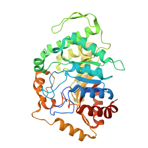 | |
UniProt | |||||
Entity Groups | |||||
| Sequence Clusters | 30% Identity50% Identity70% Identity90% Identity95% Identity100% Identity | ||||
| UniProt Group | Q0SFV5 | ||||
Sequence AnnotationsExpand | |||||
Reference Sequence | |||||
| Length ( Å ) | Angle ( ˚ ) |
|---|---|
| a = 56.859 | α = 90 |
| b = 81.631 | β = 90 |
| c = 299.15 | γ = 90 |
| Software Name | Purpose |
|---|---|
| BUSTER | refinement |
| autoPROC | data processing |
| XDS | data reduction |
| Aimless | data scaling |
| PHASER | phasing |
| Funding Organization | Location | Grant Number |
|---|---|---|
| Commonwealth Scientific and Industrial Research Organisation (CSIRO) | Australia | -- |


