8VUI | pdb_00008vui

Structure of FabS1CE-EPR-1, an elbow-locked Fab, in complex with the erythropoeitin receptor


Experimental Data Snapshot

  • Method: X-RAY DIFFRACTION
  • Resolution: 2.10 Å
  • R-Value Free: 
    0.240 (Depositor), 0.241 (DCC) 
  • R-Value Work: 
    0.197 (Depositor), 0.200 (DCC) 
  • R-Value Observed: 
    0.199 (Depositor) 

Starting Models: experimental
View more details

wwPDB Validation 3D Report Full Report

Validation slider image for 8VUI

Ligand Structure Quality Assessment 


This is version 1.1 of the entry. See complete history

Literature

Antigen-binding fragments with improved crystal lattice packing and enhanced conformational flexibility at the elbow region as crystallization chaperones.

Bruce, H.A.Singer, A.U.Blazer, L.L.Luu, K.Ploder, L.Pavlenco, A.Kurinov, I.Adams, J.J.Sidhu, S.S.

(2024) Protein Sci 33: e5081-e5081

  • DOI: https://doi.org/10.1002/pro.5081
  • Primary Citation Related Structures: 
    8VTP, 8VTR, 8VU1, 8VU4, 8VUA, 8VUC, 8VUI, 8VVM, 8VVO

  • PubMed Abstract: 

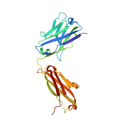
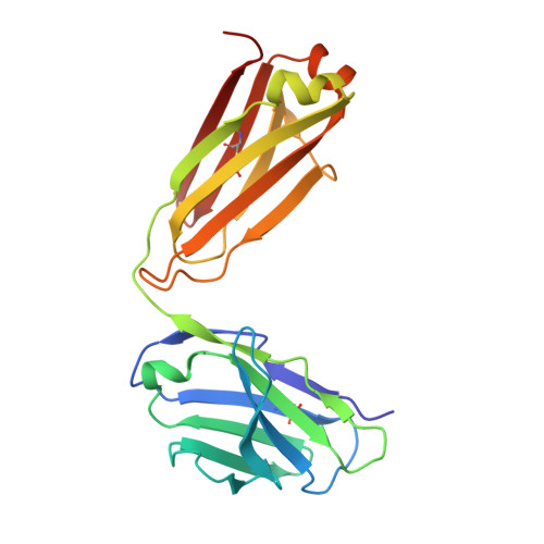
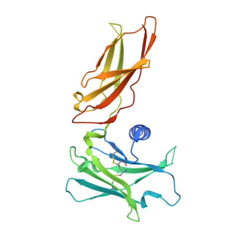
    It has been shown previously that a set of three modifications-termed S1, Crystal Kappa, and elbow-act synergistically to improve the crystallizability of an antigen-binding fragment (Fab) framework. Here, we prepared a phage-displayed library and performed crystallization screenings to identify additional substitutions-located near the heavy-chain elbow region-which cooperate with the S1, Crystal Kappa, and elbow modifications to increase expression and improve crystallizability of the Fab framework even further. One substitution (K141Q) supports the signature Crystal Kappa-mediated Fab:Fab crystal lattice packing interaction. Another substitution (E172G) improves the compatibility of the elbow modification with the Fab framework by alleviating some of the strain incurred by the shortened and bulkier elbow linker region. A third substitution (F170W) generates a split-Fab conformation, resulting in a powerful crystal lattice packing interaction comprising the biological interaction interface between the variable heavy and light chain domains. In sum, we have used K141Q, E172G, and F170W substitutions-which complement the S1, Crystal Kappa, and elbow modifications-to generate a set of highly crystallizable Fab frameworks that can be used as chaperones to enable facile elucidation of Fab:antigen complex structures by x-ray crystallography.


  • Organizational Affiliation
    • School of Pharmacy, University of Waterloo, Waterloo, Ontario, Canada.

Macromolecule Content 

  • Total Structure Weight: 73.71 kDa 
  • Atom Count: 5,016 
  • Modeled Residue Count: 626 
  • Deposited Residue Count: 668 
  • Unique protein chains: 3

Macromolecules

Find similar proteins by:|  3D Structure
Entity ID: 1
MoleculeChains  Sequence LengthOrganismDetailsImage
S1CE VARIANT OF FAB-EPR-1 heavy chain224Homo sapiensMutation(s): 0 
Find similar proteins by:|  3D Structure
Entity ID: 2
MoleculeChains  Sequence LengthOrganismDetailsImage
S1CE VARIANT OF FAB-EPR-1 light chainB [auth G]212Homo sapiensMutation(s): 0 
Find similar proteins by:|  3D Structure
Entity ID: 3
MoleculeChains  Sequence LengthOrganismDetailsImage
Erythropoietin receptorC [auth D]232Homo sapiensMutation(s): 0 
Gene Names: EPOR
UniProt & NIH Common Fund Data Resources
Find proteins for P19235 (Homo sapiens)
Explore P19235 
Go to UniProtKB:  P19235
PHAROS:  P19235
GTEx:  ENSG00000187266 
Entity Groups
Sequence Clusters30% Identity50% Identity70% Identity90% Identity95% Identity100% Identity
UniProt GroupP19235
Glycosylation
Glycosylation Sites: 1Go to GlyGen: P19235-1
Sequence Annotations
Expand
Reference Sequence

Small Molecules

Ligands 6 Unique
IDChains Name / Formula / InChI Key2D Diagram3D Interactions
NAG

Query on NAG


Download:Ideal Coordinates CCD File
N [auth D]2-acetamido-2-deoxy-beta-D-glucopyranose
C8 H15 N O6
OVRNDRQMDRJTHS-FMDGEEDCSA-N
CIT

Query on CIT


Download:Ideal Coordinates CCD File
O [auth D]CITRIC ACID
C6 H8 O7
KRKNYBCHXYNGOX-UHFFFAOYSA-N
EDO

Query on EDO


Download:Ideal Coordinates CCD File
D [auth A],
I [auth G],
J [auth G]
1,2-ETHANEDIOL
C2 H6 O2
LYCAIKOWRPUZTN-UHFFFAOYSA-N
CL

Query on CL


Download:Ideal Coordinates CCD File
G [auth A],
H [auth A],
M [auth G],
Q [auth D],
R [auth D]
CHLORIDE ION
Cl
VEXZGXHMUGYJMC-UHFFFAOYSA-M
NA

Query on NA


Download:Ideal Coordinates CCD File
E [auth A],
F [auth A],
K [auth G],
L [auth G]
SODIUM ION
Na
FKNQFGJONOIPTF-UHFFFAOYSA-N
NH4

Query on NH4


Download:Ideal Coordinates CCD File
P [auth D]AMMONIUM ION
H4 N
QGZKDVFQNNGYKY-UHFFFAOYSA-O

Experimental Data & Validation

Experimental Data

  • Method: X-RAY DIFFRACTION
  • Resolution: 2.10 Å
  • R-Value Free:  0.240 (Depositor), 0.241 (DCC) 
  • R-Value Work:  0.197 (Depositor), 0.200 (DCC) 
  • R-Value Observed: 0.199 (Depositor) 
Space Group: P 21 21 21
Unit Cell:
Length ( Å )Angle ( ˚ )
a = 56.39α = 90
b = 71.93β = 90
c = 197.02γ = 90
Software Package:
Software NamePurpose
PHENIXrefinement
MOSFLMdata reduction
Aimlessdata scaling
PHASERphasing

Structure Validation

View Full Validation Report


Ligand Structure Quality Assessment 


Entry History 

& Funding Information

Deposition Data


Funding OrganizationLocationGrant Number
Canadian Institutes of Health Research (CIHR)CanadaPJT-159640
Other privateUnited States--

Revision History  (Full details and data files)

  • Version 1.0: 2024-07-10
    Type: Initial release
  • Version 1.1: 2024-10-23
    Changes: Structure summary