8VIA | pdb_00008via

Protective effect of human non-neutralizing cross-reactive spike antibodies elicited by SARS-CoV-2 mRNA vaccination


Experimental Data Snapshot

  • Method: ELECTRON MICROSCOPY
  • Resolution: 3.60 Å
  • Aggregation State: PARTICLE 
  • Reconstruction Method: SINGLE PARTICLE 

wwPDB Validation 3D Report Full Report

Validation slider image for 8VIA

This is version 1.2 of the entry. See complete history

Literature

Protective effect and molecular mechanisms of human non-neutralizing cross-reactive spike antibodies elicited by SARS-CoV-2 mRNA vaccination.

Clark, J.J.Hoxie, I.Adelsberg, D.C.Sapse, I.A.Andreata-Santos, R.Yong, J.S.Amanat, F.Tcheou, J.Raskin, A.Singh, G.Gonzalez-Dominguez, I.Edgar, J.E.Bournazos, S.Sun, W.Carreno, J.M.Simon, V.Ellebedy, A.H.Bajic, G.Krammer, F.

(2024) Cell Rep 43: 114922-114922

  • DOI: https://doi.org/10.1016/j.celrep.2024.114922
  • Primary Citation Related Structures: 
    8VIA

  • PubMed Abstract: 

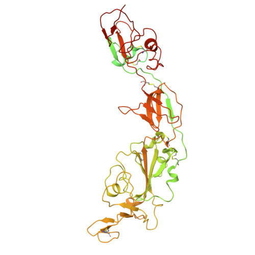
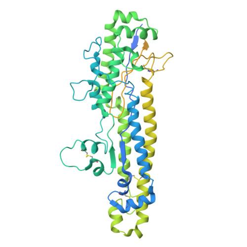
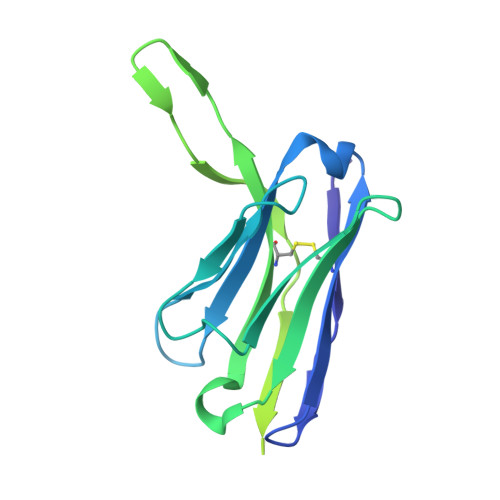
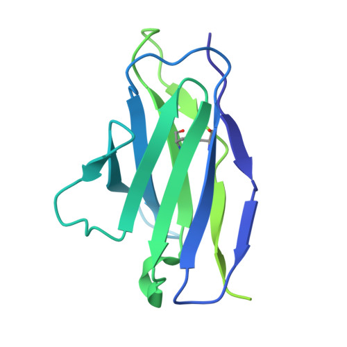
    Neutralizing antibodies correlate with protection against severe acute respiratory syndrome coronavirus 2 (SARS-CoV-2). Recent studies, however, show that binding antibody titers, in the absence of robust neutralizing activity, also correlate with protection against disease progression. Non-neutralizing antibodies cannot directly protect against infection but may recruit effector cells and thus contribute to the clearance of infected cells. Additionally, they often bind conserved epitopes across multiple variants. Here, we characterize 42 human monoclonal antibodies (mAbs) from coronavirus disease 2019 (COVID-19)-vaccinated individuals. Most of these antibodies exhibit no neutralizing activity in vitro, but several non-neutralizing antibodies provide protection against lethal challenge with SARS-CoV-2 in different animal models. A subset of those mAbs shows a clear dependence on Fc-mediated effector functions. We have determined the structures of three non-neutralizing antibodies, with two targeting the receptor-binding domain and one that binds the subdomain 1 region. Our data confirm the real-world observation in humans that non-neutralizing antibodies to SARS-CoV-2 can be protective.


  • Organizational Affiliation
    • Department of Microbiology, Icahn School of Medicine at Mount Sinai, New York, NY 10029, USA; Center for Vaccine Research and Pandemic Preparedness (C-VARPP), Icahn School of Medicine at Mount Sinai, New York, NY 10029, USA.

Macromolecule Content 

  • Total Structure Weight: 186.38 kDa 
  • Atom Count: 7,580 
  • Modeled Residue Count: 974 
  • Deposited Residue Count: 1,684 
  • Unique protein chains: 4

Macromolecules

Find similar proteins by:|  3D Structure
Entity ID: 1
MoleculeChains  Sequence LengthOrganismDetailsImage
Spike protein S1A [auth D]662Severe acute respiratory syndrome coronavirus 2Mutation(s): 0 
Gene Names: S2
UniProt
Find proteins for P0DTC2 (Severe acute respiratory syndrome coronavirus 2)
Explore P0DTC2 
Go to UniProtKB:  P0DTC2
Entity Groups
Sequence Clusters30% Identity50% Identity70% Identity90% Identity95% Identity100% Identity
UniProt GroupP0DTC2
Glycosylation
Glycosylation Sites: 3Go to GlyGen: P0DTC2-1
Sequence Annotations
Expand
Reference Sequence
Find similar proteins by:|  3D Structure
Entity ID: 2
MoleculeChains  Sequence LengthOrganismDetailsImage
Spike protein S2B [auth C]570Severe acute respiratory syndrome coronavirus 2Mutation(s): 0 
Gene Names: S2
UniProt
Find proteins for P0DTC2 (Severe acute respiratory syndrome coronavirus 2)
Explore P0DTC2 
Go to UniProtKB:  P0DTC2
Entity Groups
Sequence Clusters30% Identity50% Identity70% Identity90% Identity95% Identity100% Identity
UniProt GroupP0DTC2
Sequence Annotations
Expand
Reference Sequence
Find similar proteins by:|  3D Structure
Entity ID: 3
MoleculeChains  Sequence LengthOrganismDetailsImage
PVI.V5-4 heavy chainC [auth H]236Homo sapiensMutation(s): 0 
Find similar proteins by:|  3D Structure
Entity ID: 4
MoleculeChains  Sequence LengthOrganismDetailsImage
PVI.V5-4 light chainD [auth L]216Homo sapiensMutation(s): 0 

Oligosaccharides

Help  
Entity ID: 5
MoleculeChains Length2D Diagram GlycosylationD Interactions
2-acetamido-2-deoxy-beta-D-glucopyranose-(1-4)-2-acetamido-2-deoxy-beta-D-glucopyranoseE [auth A]2N-Glycosylation
Glycosylation Resources
GlyTouCan: G42666HT
GlyCosmos: G42666HT
GlyGen: G42666HT

Experimental Data & Validation

Experimental Data

  • Method: ELECTRON MICROSCOPY
  • Resolution: 3.60 Å
  • Aggregation State: PARTICLE 
  • Reconstruction Method: SINGLE PARTICLE 

Structure Validation

View Full Validation Report


Entry History 

& Funding Information

Deposition Data

  • Released Date: 2024-10-23 
  • Deposition Author(s): Bajic, G.

Funding OrganizationLocationGrant Number
National Institutes of Health/National Institute Of Allergy and Infectious Diseases (NIH/NIAID)United StatesAI168178

Revision History  (Full details and data files)

  • Version 1.0: 2024-10-23
    Type: Initial release
  • Version 1.1: 2024-11-06
    Changes: Data collection
  • Version 1.2: 2024-11-27
    Changes: Data collection, Database references