8V5V | pdb_00008v5v

Structure of a SARS-CoV-2 spike S2 subunit in a pre-fusion, open conformation


Experimental Data Snapshot

  • Method: ELECTRON MICROSCOPY
  • Resolution: 2.93 Å
  • Aggregation State: PARTICLE 
  • Reconstruction Method: SINGLE PARTICLE 

Starting Model: experimental
View more details

wwPDB Validation 3D Report Full Report

Validation slider image for 8V5V

This is version 1.1 of the entry. See complete history

Literature

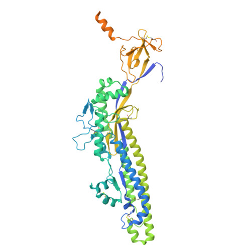
Structure of a SARS-CoV-2 spike S2 subunit in a pre-fusion, open conformation.

Olmedillas, E.Rajamanickam, R.R.Avalos, R.D.Ana-Sosa-Batiz, F.Zyla, D.Zandonatti, M.A.Harkins, S.S.Shresta, S.Hastie, K.M.Saphire, E.O.

(2025) Cell Rep 44: 116052-116052

  • DOI: https://doi.org/10.1016/j.celrep.2025.116052
  • Primary Citation Related Structures: 
    8V5V

  • PubMed Abstract: 

    The continued emergence of SARS-CoV-2 variants necessitates the development of immunogens that promote broad and durable immunity. The SARS-CoV-2 S2 fusion subunit drives viral entry and has sequence conservation among coronavirus spike proteins. Therefore, S2 could represent an immunogen to boost broadly reactive antibodies. However, when expressed without the S1 domain, metastable S2 irreversibly collapses into the post-fusion six-helix bundle conformation. Beyond well-characterized RBD/NTD shifts, biophysical measurements indicate that spike exhibits reversible "breathing" motions. Using an engineered S2-only antigen that retains the pre-fusion viral surface conformation, we isolated S2-specific antibodies from convalescent and vaccinated individuals. One mAb was used to solve a high-resolution cryo-EM structure of pre-fusion S2. Our structure reveals that, relative to intact spike, engineered S2 adopts a more "open" conformation with stabilizing intermolecular interactions at the trimer base and fusion peptide repositioning. This structure could advance next-generation "booster" immunogens and illuminate potential breathing adjustments of the coronavirus spike.


  • Organizational Affiliation
    • Center for Vaccine Innovation, La Jolla Institute for Immunology, La Jolla, CA 92037, USA.

Macromolecule Content 

  • Total Structure Weight: 274.3 kDa 
  • Atom Count: 15,882 
  • Modeled Residue Count: 2,025 
  • Deposited Residue Count: 2,463 
  • Unique protein chains: 3

Macromolecules

Find similar proteins by:|  3D Structure
Entity ID: 1
MoleculeChains  Sequence LengthOrganismDetailsImage
Spike protein S2,FibritinA [auth G],
B [auth E],
C [auth F]
588Severe acute respiratory syndrome coronavirus 2Tequatrovirus T4
This entity is chimeric
Mutation(s): 8 
Gene Names: S2wac
UniProt
Find proteins for P0DTC2 (Severe acute respiratory syndrome coronavirus 2)
Explore P0DTC2 
Go to UniProtKB:  P0DTC2
Find proteins for P10104 (Enterobacteria phage T4)
Explore P10104 
Go to UniProtKB:  P10104
Entity Groups
Sequence Clusters30% Identity50% Identity70% Identity90% Identity95% Identity100% Identity
UniProt GroupsP0DTC2P10104
Glycosylation
Glycosylation Sites: 6Go to GlyGen: P0DTC2-1
Sequence Annotations
Expand
Reference Sequence
Find similar proteins by:|  3D Structure
Entity ID: 2
MoleculeChains  Sequence LengthOrganismDetailsImage
Variable domain of the light chain from the human antibody 6C10D [auth d],
E [auth g],
H [auth e]
108Homo sapiensMutation(s): 0 
Find similar proteins by:|  3D Structure
Entity ID: 3
MoleculeChains  Sequence LengthOrganismDetailsImage
Variable domain of the heavy chain from the human antibody 6C10F [auth f],
G [auth c],
I [auth b]
125Homo sapiensMutation(s): 0 

Oligosaccharides

Help  
Entity ID: 4
MoleculeChains Length2D Diagram GlycosylationD Interactions
2-acetamido-2-deoxy-beta-D-glucopyranose-(1-4)-2-acetamido-2-deoxy-beta-D-glucopyranoseJ [auth A],
K [auth B],
L [auth C]
2N-Glycosylation
Glycosylation Resources
GlyTouCan: G42666HT
GlyCosmos: G42666HT
GlyGen: G42666HT

Small Molecules

Ligands 1 Unique
IDChains Name / Formula / InChI Key2D Diagram3D Interactions
NAG

Query on NAG


Download:Ideal Coordinates CCD File
AA [auth F]
M [auth G]
N [auth G]
O [auth G]
P [auth G]
AA [auth F],
M [auth G],
N [auth G],
O [auth G],
P [auth G],
Q [auth G],
R [auth E],
S [auth E],
T [auth E],
U [auth E],
V [auth E],
W [auth F],
X [auth F],
Y [auth F],
Z [auth F]
2-acetamido-2-deoxy-beta-D-glucopyranose
C8 H15 N O6
OVRNDRQMDRJTHS-FMDGEEDCSA-N

Experimental Data & Validation

Experimental Data

  • Method: ELECTRON MICROSCOPY
  • Resolution: 2.93 Å
  • Aggregation State: PARTICLE 
  • Reconstruction Method: SINGLE PARTICLE 
EM Software:
TaskSoftware PackageVersion
RECONSTRUCTIONcryoSPARC4.0
MODEL REFINEMENTPHENIX1.20.1_4487:1.21
MODEL REFINEMENTCoot0.9.5

Structure Validation

View Full Validation Report


Entry History 

& Funding Information

Deposition Data


Funding OrganizationLocationGrant Number
National Institutes of Health/National Institute Of Allergy and Infectious Diseases (NIH/NIAID)United StatesAI165072

Revision History  (Full details and data files)

  • Version 1.0: 2024-12-18
    Type: Initial release
  • Version 1.1: 2025-08-06
    Changes: Data collection, Database references