Identification of small molecule inhibitors of G3BP-driven stress granule formation.
Freibaum, B.D., Messing, J., Nakamura, H., Yurtsever, U., Wu, J., Kim, H.J., Hixon, J., Lemieux, R.M., Duffner, J., Huynh, W., Wong, K., White, M., Lee, C., Meyers, R.E., Parker, R., Taylor, J.P.(2024) J Cell Biol 223
- PubMed: 38284934 Search on PubMedSearch on PubMed Central
- DOI: https://doi.org/10.1083/jcb.202308083
- Primary Citation Related Structures:
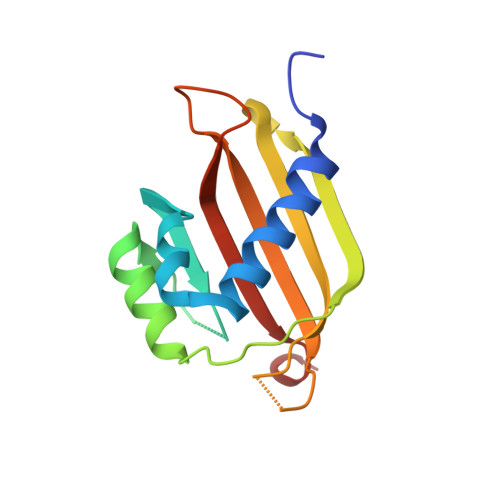
8V1L - PubMed Abstract:
Stress granule formation is triggered by the release of mRNAs from polysomes and is promoted by the action of the RNA-binding proteins G3BP1/2. Stress granules have been implicated in several disease states, including cancer and neurodegeneration. Consequently, compounds that limit stress granule formation or promote their dissolution have potential as both experimental tools and novel therapeutics. Herein, we describe two small molecules, G3BP inhibitor a and b (G3Ia and G3Ib), designed to bind to a specific pocket in G3BP1/2 that is targeted by viral inhibitors of G3BP1/2 function. In addition to disrupting the co-condensation of RNA, G3BP1, and caprin 1 in vitro, these compounds inhibit stress granule formation in cells treated prior to or concurrent with stress and dissolve pre-existing stress granules. These effects are consistent across multiple cell types and a variety of initiating stressors. Thus, these compounds represent powerful tools to probe the biology of stress granules and hold promise for therapeutic interventions designed to modulate stress granule formation.
- Department of Cell and Molecular Biology, St. Jude Children's Research Hospital, Memphis, TN, USA.
Organizational Affiliation: