Cryo-EM structure of bacterial nitrilase reveals insight into oligomerization, substrate recognition, and catalysis.
Aguirre-Sampieri, S., Casanal, A., Emsley, P., Garza-Ramos, G.(2024) J Struct Biol 216: 108093-108093
- PubMed: 38615726 Search on PubMed
- DOI: https://doi.org/10.1016/j.jsb.2024.108093
- Primary Citation Related Structures:
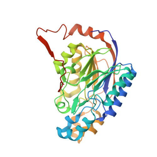
8UXU - PubMed Abstract:
Many enzymes can self-assemble into higher-order structures with helical symmetry. A particularly noteworthy example is that of nitrilases, enzymes in which oligomerization of dimers into spiral homo-oligomers is a requirement for their enzymatic function. Nitrilases are widespread in nature where they catalyze the hydrolysis of nitriles into the corresponding carboxylic acid and ammonia. Here, we present the Cryo-EM structure, at 3 Å resolution, of a C-terminal truncate nitrilase from Rhodococcus sp. V51B that assembles in helical filaments. The model comprises a complete turn of the helical arrangement with a substrate-intermediate bound to the catalytic cysteine. The structure was solved having added the substrate to the protein. The length and stability of filaments was made more substantial in the presence of the aromatic substrate, benzonitrile, but not for aliphatic nitriles or dinitriles. The overall structure maintains the topology of the nitrilase family, and the filament is formed by the association of dimers in a chain-like mechanism that stabilizes the spiral. The active site is completely buried inside each monomer, while the substrate binding pocket was observed within the oligomerization interfaces. The present structure is in a closed configuration, judging by the position of the lid, suggesting that the intermediate is one of the covalent adducts. The proximity of the active site to the dimerization and oligomerization interfaces, allows the dimer to sense structural changes once the benzonitrile was bound, and translated to the rest of the filament, stabilizing the helical structure.
- Universidad Nacional Autónoma de México, Facultad de Medicina, Departamento de Bioquímica, Circuito Escolar S/N, Ciudad Universitaria, CDMX, Mexico.
Organizational Affiliation: