Highly Similar Tetramerization Domains from the p53 Protein of Different Mammalian Species Possess Varying Biophysical, Functional and Structural Properties.
Sakaguchi, S., Nakagawa, N., Wahba, H.M., Wada, J., Kamada, R., Omichinski, J.G., Sakaguchi, K.(2023) Int J Mol Sci 24
- PubMed: 38068946 Search on PubMedSearch on PubMed Central
- DOI: https://doi.org/10.3390/ijms242316620
- Primary Citation Related Structures:
8UQR, 8UQS, 8UQT - PubMed Abstract:
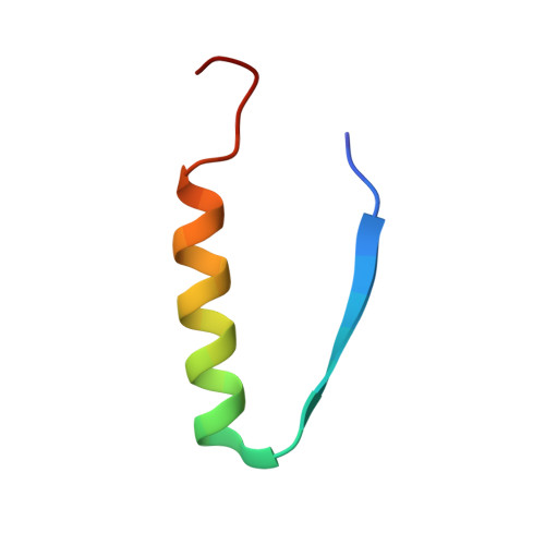
The p53 protein is a transcriptional regulatory factor and many of its functions require that it forms a tetrameric structure. Although the tetramerization domain of mammalian p53 proteins (p53TD) share significant sequence similarities, it was recently shown that the tree shrew p53TD is considerably more thermostable than the human p53TD. To determine whether other mammalian species display differences in this domain, we used biophysical, functional, and structural studies to compare the properties of the p53TDs from six mammalian model organisms (human, tree shrew, guinea pig, Chinese hamster, sheep, and opossum). The results indicate that the p53TD from the opossum and tree shrew are significantly more stable than the human p53TD, and there is a correlation between the thermostability of the p53TDs and their ability to activate transcription. Structural analysis of the tree shrew and opossum p53TDs indicated that amino acid substitutions within two distinct regions of their p53TDs can dramatically alter hydrophobic packing of the tetramer, and in particular substitutions at positions corresponding to F341 and Q354 of the human p53TD. Together, the results suggest that subtle changes in the sequence of the p53TD can dramatically alter the stability, and potentially lead to important changes in the functional activity, of the p53 protein.
- Department of Chemistry, Faculty of Science, Hokkaido University, Sapporo 060-0810, Japan.
Organizational Affiliation: