


Streptococcus surface alpha enolase exposed dimers were found to be the active form on lipid surface that binds to human plasminogen
Tjia-Fleck, S., Readnour, B.M., Castellino, F.J.To be published.
Experimental Data Snapshot
wwPDB Validation 3D Report Full Report

Entity ID: 1 | |||||
|---|---|---|---|---|---|
| Molecule | Chains | Sequence Length | Organism | Details | Image |
| Enolase | 436 | Streptococcus pyogenes | Mutation(s): 0 Gene Names: eno, E0F66_01300, E0F67_02490, FGO82_08975, SAMEA1711581_01820, SAMEA864267_00487 EC: 4.2.1.11 | 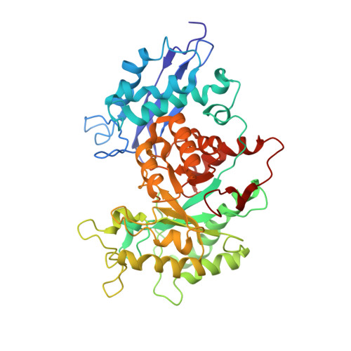 | |
UniProt | |||||
Find proteins for P69949 (Streptococcus pyogenes serotype M1) Explore P69949 Go to UniProtKB: P69949 | |||||
Entity Groups | |||||
| Sequence Clusters | 30% Identity50% Identity70% Identity90% Identity95% Identity100% Identity | ||||
| UniProt Group | P69949 | ||||
Sequence AnnotationsExpand | |||||
| |||||
| Task | Software Package | Version |
|---|---|---|
| RECONSTRUCTION | UCSF ChimeraX |
| Funding Organization | Location | Grant Number |
|---|---|---|
| National Institutes of Health/National Heart, Lung, and Blood Institute (NIH/NHLBI) | United States | HL013423 |


