8UFU

Structure of human endothelial nitric oxide synthase heme domain in complex with 7-(9-amino-6,7,8,9-tetrahydro-5H-benzo[7]annulen-2-yl)-4-methylquinolin-2-amine


Experimental Data Snapshot

  • Method: X-RAY DIFFRACTION
  • Resolution: 2.05 Å
  • R-Value Free: 0.256 
  • R-Value Work: 0.205 
  • R-Value Observed: 0.207 

wwPDB Validation   3D Report Full Report


Ligand Structure Quality Assessment 


This is version 1.1 of the entry. See complete history


Literature

Crystallographic and Computational Insights into Isoform-Selective Dynamics in Nitric Oxide Synthase.

Li, H.Hardy, C.D.Reidl, C.T.Jing, Q.Xue, F.Cinelli, M.Silverman, R.B.Poulos, T.L.

(2024) Biochemistry 63: 788-796

  • DOI: https://doi.org/10.1021/acs.biochem.3c00601
  • Primary Citation of Related Structures:  
    8UFP, 8UFQ, 8UFR, 8UFS, 8UFT, 8UFU

  • PubMed Abstract: 

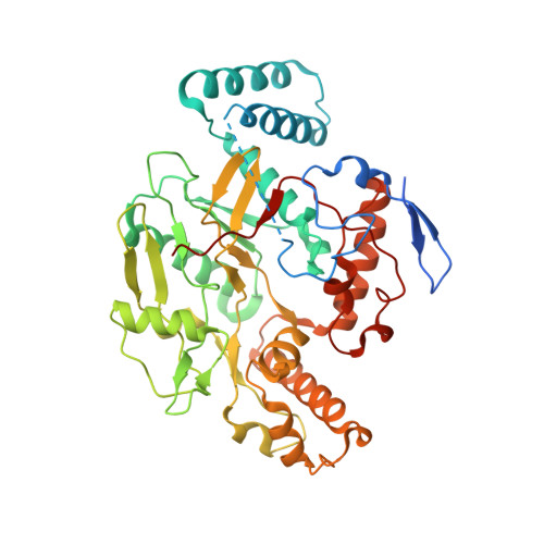
    In our efforts to develop inhibitors selective for neuronal nitric oxide synthase (nNOS) over endothelial nitric oxide synthase (eNOS), we found that nNOS can undergo conformational changes in response to inhibitor binding that does not readily occur in eNOS. One change involves movement of a conserved tyrosine, which hydrogen bonds to one of the heme propionates, but in the presence of an inhibitor, changes conformation, enabling part of the inhibitor to hydrogen bond with the heme propionate. This movement does not occur as readily in eNOS and may account for the reason why these inhibitors bind more tightly to nNOS. A second structural change occurs upon the binding of a second inhibitor molecule to nNOS, displacing the pterin cofactor. Binding of this second site inhibitor requires structural changes at the dimer interface, which also occurs more readily in nNOS than in eNOS. Here, we used a combination of crystallography, mutagenesis, and computational methods to better understand the structural basis for these differences in NOS inhibitor binding. Computational results show that a conserved tyrosine near the primary inhibitor binding site is anchored more tightly in eNOS than in nNOS, allowing for less flexibility of this residue. We also find that the inefficiency of eNOS to bind a second inhibitor molecule is likely due to the tighter dimer interface in eNOS compared with nNOS. This study provides a better understanding of how subtle structural differences in NOS isoforms can result in substantial dynamic differences that can be exploited in the development of isoform-selective inhibitors.


  • Organizational Affiliation

    Departments of Molecular Biology and Biochemistry, Pharmaceutical Sciences, and Chemistry, University of California, Irvine, California 92697-3900, United States.


Macromolecules
Find similar proteins by:  (by identity cutoff)  |  3D Structure
Entity ID: 1
MoleculeChains Sequence LengthOrganismDetailsImage
Nitric oxide synthase, endothelial
A, B, C, D
440Homo sapiensMutation(s): 1 
Gene Names: NOS3
EC: 1.14.13.39
UniProt & NIH Common Fund Data Resources
Find proteins for P29474 (Homo sapiens)
Explore P29474 
Go to UniProtKB:  P29474
PHAROS:  P29474
GTEx:  ENSG00000164867 
Entity Groups  
Sequence Clusters30% Identity50% Identity70% Identity90% Identity95% Identity100% Identity
UniProt GroupP29474
Sequence Annotations
Expand
  • Reference Sequence
Small Molecules
Ligands 8 Unique
IDChains Name / Formula / InChI Key2D Diagram3D Interactions
HEM
Query on HEM

Download Ideal Coordinates CCD File 
E [auth A],
IA [auth D],
O [auth B],
W [auth C]
PROTOPORPHYRIN IX CONTAINING FE
C34 H32 Fe N4 O4
KABFMIBPWCXCRK-RGGAHWMASA-L
WRI (Subject of Investigation/LOI)
Query on WRI

Download Ideal Coordinates CCD File 
F [auth A]
FA [auth C]
G [auth A]
JA [auth D]
P [auth B]
F [auth A],
FA [auth C],
G [auth A],
JA [auth D],
P [auth B],
Q [auth B],
X [auth C],
Y [auth C]
(7M)-7-[(9S)-9-amino-6,7,8,9-tetrahydro-5H-benzo[7]annulen-2-yl]-4-methylquinolin-2-amine
C21 H23 N3
GORDHKJSCQTIOT-IBGZPJMESA-N
BTB
Query on BTB

Download Ideal Coordinates CCD File 
AA [auth C]
H [auth A]
I [auth A]
KA [auth D]
LA [auth D]
AA [auth C],
H [auth A],
I [auth A],
KA [auth D],
LA [auth D],
R [auth B],
S [auth B],
Z [auth C]
2-[BIS-(2-HYDROXY-ETHYL)-AMINO]-2-HYDROXYMETHYL-PROPANE-1,3-DIOL
C8 H19 N O5
OWMVSZAMULFTJU-UHFFFAOYSA-N
GD
Query on GD

Download Ideal Coordinates CCD File 
OA [auth D],
U [auth B]
GADOLINIUM ATOM
Gd
UIWYJDYFSGRHKR-UHFFFAOYSA-N
GOL
Query on GOL

Download Ideal Coordinates CCD File 
BA [auth C],
J [auth A],
MA [auth D]
GLYCEROL
C3 H8 O3
PEDCQBHIVMGVHV-UHFFFAOYSA-N
ZN
Query on ZN

Download Ideal Coordinates CCD File 
DA [auth C]
EA [auth C]
GA [auth C]
L [auth A]
M [auth A]
DA [auth C],
EA [auth C],
GA [auth C],
L [auth A],
M [auth A],
V [auth B]
ZINC ION
Zn
PTFCDOFLOPIGGS-UHFFFAOYSA-N
CA
Query on CA

Download Ideal Coordinates CCD File 
HA [auth C],
N [auth A]
CALCIUM ION
Ca
BHPQYMZQTOCNFJ-UHFFFAOYSA-N
CL
Query on CL

Download Ideal Coordinates CCD File 
CA [auth C],
K [auth A],
NA [auth D],
T [auth B]
CHLORIDE ION
Cl
VEXZGXHMUGYJMC-UHFFFAOYSA-M
Experimental Data & Validation

Experimental Data

  • Method: X-RAY DIFFRACTION
  • Resolution: 2.05 Å
  • R-Value Free: 0.256 
  • R-Value Work: 0.205 
  • R-Value Observed: 0.207 
  • Space Group: P 1 21 1
Unit Cell:
Length ( Å )Angle ( ˚ )
a = 59.673α = 90
b = 152.827β = 90.82
c = 109.162γ = 90
Software Package:
Software NamePurpose
PHENIXrefinement
Aimlessdata scaling
PDB_EXTRACTdata extraction
XDSdata reduction
REFMACphasing

Structure Validation

View Full Validation Report


Ligand Structure Quality Assessment 


Entry History & Funding Information

Deposition Data


Funding OrganizationLocationGrant Number
National Institutes of Health/National Institute of General Medical Sciences (NIH/NIGMS)United StatesGM57353

Revision History  (Full details and data files)

  • Version 1.0: 2024-03-13
    Type: Initial release
  • Version 1.1: 2024-04-03
    Changes: Database references