8TWI

Cryo-EM structure of the PP2A:B55-FAM122A complex, PP2Ac body


Experimental Data Snapshot

  • Method: ELECTRON MICROSCOPY
  • Resolution: 2.69 Å
  • Aggregation State: PARTICLE 
  • Reconstruction Method: SINGLE PARTICLE 

wwPDB Validation   3D Report Full Report


This is version 1.3 of the entry. See complete history


Literature

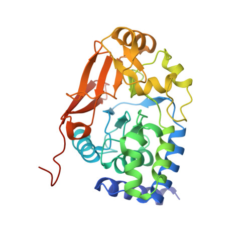
Cryo-EM structures of PP2A:B55-FAM122A and PP2A:B55-ARPP19.

Padi, S.K.R.Vos, M.R.Godek, R.J.Fuller, J.R.Kruse, T.Hein, J.B.Nilsson, J.Kelker, M.S.Page, R.Peti, W.

(2024) Nature 625: 195-203

  • DOI: https://doi.org/10.1038/s41586-023-06870-3
  • Primary Citation of Related Structures:  
    8SO0, 8TTB, 8TWE, 8TWI

  • PubMed Abstract: 

    Progression through the cell cycle is controlled by regulated and abrupt changes in phosphorylation 1 . Mitotic entry is initiated by increased phosphorylation of mitotic proteins, a process driven by kinases 2 , whereas mitotic exit is achieved by counteracting dephosphorylation, a process driven by phosphatases, especially PP2A:B55 3 . Although the role of kinases in mitotic entry is well established, recent data have shown that mitosis is only successfully initiated when the counterbalancing phosphatases are also inhibited 4 . Inhibition of PP2A:B55 is achieved by the intrinsically disordered proteins ARPP19 5,6 and FAM122A 7 . Despite their critical roles in mitosis, the mechanisms by which they achieve PP2A:B55 inhibition is unknown. Here, we report the single-particle cryo-electron microscopy structures of PP2A:B55 bound to phosphorylated ARPP19 and FAM122A. Consistent with our complementary NMR spectroscopy studies, both intrinsically disordered proteins bind PP2A:B55, but do so in highly distinct manners, leveraging multiple distinct binding sites on B55. Our extensive structural, biophysical and biochemical data explain how substrates and inhibitors are recruited to PP2A:B55 and provide a molecular roadmap for the development of therapeutic interventions for PP2A:B55-related diseases.


  • Organizational Affiliation

    Department of Molecular Biology and Biophysics, University of Connecticut Health Center, Farmington, CT, USA.


Macromolecules
Find similar proteins by:  (by identity cutoff)  |  3D Structure
Entity ID: 1
MoleculeChains Sequence LengthOrganismDetailsImage
Serine/threonine-protein phosphatase 2A 65 kDa regulatory subunit A alpha isoform584Homo sapiensMutation(s): 0 
Gene Names: PPP2R1A
UniProt & NIH Common Fund Data Resources
Find proteins for P30153 (Homo sapiens)
Explore P30153 
Go to UniProtKB:  P30153
PHAROS:  P30153
GTEx:  ENSG00000105568 
Entity Groups  
Sequence Clusters30% Identity50% Identity70% Identity90% Identity95% Identity100% Identity
UniProt GroupP30153
Sequence Annotations
Expand
  • Reference Sequence
Find similar proteins by:  (by identity cutoff)  |  3D Structure
Entity ID: 2
MoleculeChains Sequence LengthOrganismDetailsImage
Serine/threonine-protein phosphatase 2A catalytic subunit alpha isoformB [auth C]311Homo sapiensMutation(s): 0 
Gene Names: PPP2CA
EC: 3.1.3.16
UniProt & NIH Common Fund Data Resources
Find proteins for P67775 (Homo sapiens)
Explore P67775 
Go to UniProtKB:  P67775
PHAROS:  P67775
GTEx:  ENSG00000113575 
Entity Groups  
Sequence Clusters30% Identity50% Identity70% Identity90% Identity95% Identity100% Identity
UniProt GroupP67775
Sequence Annotations
Expand
  • Reference Sequence
Find similar proteins by:  (by identity cutoff)  |  3D Structure
Entity ID: 3
MoleculeChains Sequence LengthOrganismDetailsImage
PPP2R1A-PPP2R2A-interacting phosphatase regulator 1C [auth D]95Homo sapiensMutation(s): 0 
Gene Names: PABIR1C9orf42FAM122A
UniProt & NIH Common Fund Data Resources
Find proteins for Q96E09 (Homo sapiens)
Explore Q96E09 
Go to UniProtKB:  Q96E09
PHAROS:  Q96E09
GTEx:  ENSG00000187866 
Entity Groups  
Sequence Clusters30% Identity50% Identity70% Identity90% Identity95% Identity100% Identity
UniProt GroupQ96E09
Sequence Annotations
Expand
  • Reference Sequence
Small Molecules
Modified Residues  1 Unique
IDChains TypeFormula2D DiagramParent
MLL
Query on MLL
B [auth C]L-PEPTIDE LINKINGC7 H15 N O2LEU
Experimental Data & Validation

Experimental Data

  • Method: ELECTRON MICROSCOPY
  • Resolution: 2.69 Å
  • Aggregation State: PARTICLE 
  • Reconstruction Method: SINGLE PARTICLE 
EM Software:
TaskSoftware PackageVersion
MODEL REFINEMENTPHENIX1.20.1-4487
MODEL REFINEMENTCoot0.9
RECONSTRUCTIONRELION4.0

Structure Validation

View Full Validation Report


Entry History & Funding Information

Deposition Data


Funding OrganizationLocationGrant Number
National Institutes of Health/National Institute of General Medical Sciences (NIH/NIGMS)United StatesR01GM144379
National Institutes of Health/National Institute of General Medical Sciences (NIH/NIGMS)United StatesR01GM134683
National Institutes of Health/National Institute of Neurological Disorders and Stroke (NIH/NINDS)United StatesR01NS124666

Revision History  (Full details and data files)

  • Version 1.0: 2023-11-01
    Type: Initial release
  • Version 1.1: 2023-11-15
    Changes: Data collection
  • Version 1.2: 2024-01-10
    Changes: Database references
  • Version 1.3: 2024-01-17
    Changes: Database references