8T2H | pdb_00008t2h

DYRK1A complex with DYR530


Experimental Data Snapshot

  • Method: X-RAY DIFFRACTION
  • Resolution: 1.85 Å
  • R-Value Free: 
    0.213 (Depositor), 0.223 (DCC) 
  • R-Value Work: 
    0.163 (Depositor), 0.198 (DCC) 
  • R-Value Observed: 
    0.166 (Depositor) 

Starting Model: experimental
View more details

wwPDB Validation 3D Report Full Report

Validation slider image for 8T2H

Ligand Structure Quality Assessment 


This is version 1.2 of the entry. See complete history

Literature

Discovery and Functional Characterization of a Potent, Selective, and Metabolically Stable PROTAC of the Protein Kinases DYRK1A and DYRK1B.

Wilms, G.Schofield, K.Maddern, S.Foley, C.Shaw, Y.Smith, B.Basantes, L.E.Schwandt, K.Babendreyer, A.Chavez, T.McKee, N.Gokhale, V.Kallabis, S.Meissner, F.Rokey, S.N.Dunckley, T.Montfort, W.R.Becker, W.Hulme, C.

(2024) J Med Chem 67: 17259-17289

  • DOI: https://doi.org/10.1021/acs.jmedchem.4c01130
  • Primary Citation Related Structures: 
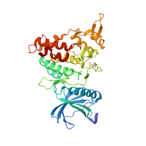
    8T2H

  • PubMed Abstract: 

    Small-molecule-induced protein degradation has emerged as a promising pharmacological modality for inactivating disease-relevant protein kinases. DYRK1A and DYRK1B are closely related protein kinases that are involved in pathological processes such as neurodegeneration, cancer development, and adaptive immune homeostasis. Herein, we report the development of the first DYRK1 proteolysis targeting chimeras (PROTACs) that combine a new ATP-competitive DYRK1 inhibitor with ligands for the E3 ubiquitin ligase component cereblon (CRBN) to induce ubiquitination and subsequent proteasomal degradation of DYRK1A and DYRK1B. The lead compound (DYR684) promoted fast, efficient, potent, and selective degradation of DYRK1A in cell-based assays. Interestingly, an enzymatically inactive splicing variant of DYRK1B (p65) resisted degradation. Compared to competitive kinase inhibition, targeted degradation of DYRK1 by DYR684 provided improved suppression of downstream signaling. Collectively, our results identify DYRKs as viable targets for PROTAC-mediated degradation and qualify DYR684 as a useful chemical probe for DYRK1A and DYRK1B.


  • Organizational Affiliation
    • Institute of Pharmacology and Toxicology, RWTH Aachen University, Wendlingweg 2, 52074 Aachen, Germany.

Macromolecule Content 

  • Total Structure Weight: 86.18 kDa 
  • Atom Count: 6,053 
  • Modeled Residue Count: 681 
  • Deposited Residue Count: 730 
  • Unique protein chains: 1

Macromolecules

Find similar proteins by:|  3D Structure
Entity ID: 1
MoleculeChains  Sequence LengthOrganismDetailsImage
Dual specificity tyrosine-phosphorylation-regulated kinase 1A
A, B
365Homo sapiensMutation(s): 0 
Gene Names: DYRK1ADYRKMNBMNBH
EC: 2.7.11.23 (PDB Primary Data), 2.7.12.1 (PDB Primary Data)
UniProt & NIH Common Fund Data Resources
Find proteins for Q13627 (Homo sapiens)
Explore Q13627 
Go to UniProtKB:  Q13627
PHAROS:  Q13627
GTEx:  ENSG00000157540 
Entity Groups
Sequence Clusters30% Identity50% Identity70% Identity90% Identity95% Identity100% Identity
UniProt GroupQ13627
Sequence Annotations
Expand
Reference Sequence

Small Molecules

Ligands 3 Unique
IDChains Name / Formula / InChI Key2D Diagram3D Interactions
XIR
(Subject of Investigation/LOI)

Query on XIR


Download:Ideal Coordinates CCD File
C [auth A],
G [auth B]
(4P)-4-{(3M)-3-[3-fluoro-4-(4-methylpiperazin-1-yl)phenyl]-2-methyl-3H-imidazo[4,5-b]pyridin-5-yl}pyridin-2-amine
C23 H24 F N7
HGZWGWAZKLVNRM-UHFFFAOYSA-N
PG4

Query on PG4


Download:Ideal Coordinates CCD File
D [auth A]TETRAETHYLENE GLYCOL
C8 H18 O5
UWHCKJMYHZGTIT-UHFFFAOYSA-N
GOL

Query on GOL


Download:Ideal Coordinates CCD File
E [auth A],
F [auth A],
H [auth B]
GLYCEROL
C3 H8 O3
PEDCQBHIVMGVHV-UHFFFAOYSA-N
Modified Residues  1 Unique
IDChains TypeFormula2D DiagramParent
PTR
Query on PTR
A, B
L-PEPTIDE LINKINGC9 H12 N O6 PTYR

Experimental Data & Validation

Experimental Data

  • Method: X-RAY DIFFRACTION
  • Resolution: 1.85 Å
  • R-Value Free:  0.213 (Depositor), 0.223 (DCC) 
  • R-Value Work:  0.163 (Depositor), 0.198 (DCC) 
  • R-Value Observed: 0.166 (Depositor) 
Space Group: P 21 21 21
Unit Cell:
Length ( Å )Angle ( ˚ )
a = 63.472α = 90
b = 83.213β = 90
c = 146.675γ = 90
Software Package:
Software NamePurpose
REFMACrefinement
PROTEUM2data reduction
Aimlessdata scaling
MOLREPphasing

Structure Validation

View Full Validation Report


Ligand Structure Quality Assessment 


Entry History 

& Funding Information

Deposition Data


Funding OrganizationLocationGrant Number
National Institutes of Health/National Institute on Aging (NIH/NIA)United StatesAG067926

Revision History  (Full details and data files)

  • Version 1.0: 2024-05-15
    Type: Initial release
  • Version 1.1: 2024-10-30
    Changes: Structure summary
  • Version 1.2: 2024-11-27
    Changes: Database references