8SOC

Phosphoinositide phosphate 3 kinase gamma bound with ADP and Gbetagamma


Experimental Data Snapshot

  • Method: ELECTRON MICROSCOPY
  • Resolution: 3.50 Å
  • Aggregation State: PARTICLE 
  • Reconstruction Method: SINGLE PARTICLE 

wwPDB Validation   3D Report Full Report


This is version 1.1 of the entry. See complete history


Literature

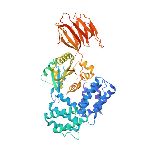
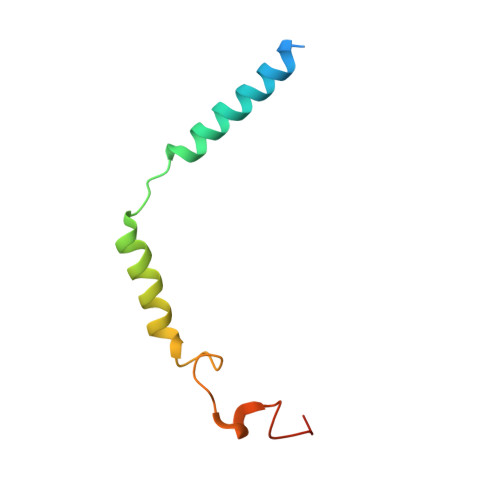
Molecular basis for G beta gamma-mediated activation of phosphoinositide 3-kinase gamma.

Chen, C.L.Syahirah, R.Ravala, S.K.Yen, Y.C.Klose, T.Deng, Q.Tesmer, J.J.G.

(2024) Nat Struct Mol Biol 

  • DOI: https://doi.org/10.1038/s41594-024-01265-y
  • Primary Citation of Related Structures:  
    8SO9, 8SOA, 8SOB, 8SOC, 8SOD, 8SOE

  • PubMed Abstract: 

    The conversion of phosphatidylinositol 4,5-bisphosphate to phosphatidylinositol 3,4,5-triphosphate by phosphoinositide 3-kinase γ (PI3Kγ) is critical for neutrophil chemotaxis and cancer metastasis. PI3Kγ is activated by Gβγ heterodimers released from G protein-coupled receptors responding to extracellular signals. Here we determined cryo-electron microscopy structures of Sus scrofa PI3Kγ-human Gβγ complexes in the presence of substrates/analogs, revealing two Gβγ binding sites: one on the p110γ helical domain and another on the p101 C-terminal domain. Comparison with PI3Kγ alone reveals conformational changes in the kinase domain upon Gβγ binding that are similar to Ras·GTP-induced changes. Assays of variants perturbing the Gβγ binding sites and interdomain contacts altered by Gβγ binding suggest that Gβγ recruits the enzyme to membranes and allosterically regulates activity via both sites. Studies of zebrafish neutrophil migration align with these findings, paving the way for in-depth investigation of Gβγ-mediated activation mechanisms in this enzyme family and drug development for PI3Kγ.


  • Organizational Affiliation

    Department of Biological Sciences, Purdue University, West Lafayette, IN, USA.


Macromolecules
Find similar proteins by:  (by identity cutoff)  |  3D Structure
Entity ID: 1
MoleculeChains Sequence LengthOrganismDetailsImage
phosphatidylinositol-4,5-bisphosphate 3-kinase1,108Sus scrofaMutation(s): 0 
UniProt
Find proteins for A0A8D1WUA4 (Sus scrofa)
Explore A0A8D1WUA4 
Go to UniProtKB:  A0A8D1WUA4
Entity Groups  
Sequence Clusters30% Identity50% Identity70% Identity90% Identity95% Identity100% Identity
UniProt GroupA0A8D1WUA4
Sequence Annotations
Expand
  • Reference Sequence
Find similar proteins by:  (by identity cutoff)  |  3D Structure
Entity ID: 2
MoleculeChains Sequence LengthOrganismDetailsImage
Phosphoinositide 3-kinase regulatory subunit 5890Sus scrofaMutation(s): 0 
UniProt
Find proteins for A0A8D0T2D6 (Sus scrofa)
Explore A0A8D0T2D6 
Go to UniProtKB:  A0A8D0T2D6
Entity Groups  
Sequence Clusters30% Identity50% Identity70% Identity90% Identity95% Identity100% Identity
UniProt GroupA0A8D0T2D6
Sequence Annotations
Expand
  • Reference Sequence
Find similar proteins by:  (by identity cutoff)  |  3D Structure
Entity ID: 3
MoleculeChains Sequence LengthOrganismDetailsImage
Guanine nucleotide-binding protein G(I)/G(S)/G(T) subunit beta-1340Bos taurusMutation(s): 0 
Gene Names: GNB1
UniProt
Find proteins for P62871 (Bos taurus)
Explore P62871 
Go to UniProtKB:  P62871
Entity Groups  
Sequence Clusters30% Identity50% Identity70% Identity90% Identity95% Identity100% Identity
UniProt GroupP62871
Sequence Annotations
Expand
  • Reference Sequence
Find similar proteins by:  (by identity cutoff)  |  3D Structure
Entity ID: 4
MoleculeChains Sequence LengthOrganismDetailsImage
Guanine nucleotide-binding protein G(I)/G(S)/G(O) subunit gamma-2D [auth G]77Bos taurusMutation(s): 1 
Gene Names: GNG2
UniProt
Find proteins for P63212 (Bos taurus)
Explore P63212 
Go to UniProtKB:  P63212
Entity Groups  
Sequence Clusters30% Identity50% Identity70% Identity90% Identity95% Identity100% Identity
UniProt GroupP63212
Sequence Annotations
Expand
  • Reference Sequence
Small Molecules
Ligands 1 Unique
IDChains Name / Formula / InChI Key2D Diagram3D Interactions
ADP (Subject of Investigation/LOI)
Query on ADP

Download Ideal Coordinates CCD File 
E [auth A]ADENOSINE-5'-DIPHOSPHATE
C10 H15 N5 O10 P2
XTWYTFMLZFPYCI-KQYNXXCUSA-N
Experimental Data & Validation

Experimental Data

  • Method: ELECTRON MICROSCOPY
  • Resolution: 3.50 Å
  • Aggregation State: PARTICLE 
  • Reconstruction Method: SINGLE PARTICLE 
EM Software:
TaskSoftware PackageVersion
RECONSTRUCTIONcryoSPARC3.3.1
MODEL REFINEMENTCoot9.6
MODEL REFINEMENTPHENIX1.20-4459

Structure Validation

View Full Validation Report


Entry History & Funding Information

Deposition Data


Funding OrganizationLocationGrant Number
American Heart AssociationUnited States834497

Revision History  (Full details and data files)

  • Version 1.0: 2024-04-03
    Type: Initial release
  • Version 1.1: 2024-04-17
    Changes: Database references