8SDK

The MicroED structure of proteinase K crystallized by suspended drop crystallization

  • Classification: HYDROLASE
  • Organism(s): Parengyodontium album
  • Mutation(s): No 

  • Deposited: 2023-04-06 Released: 2023-05-31 
  • Deposition Author(s): Gillman, C., Nicolas, W.J., Martynowycz, M.W., Gonen, T.
  • Funding Organization(s): National Institutes of Health/National Institute of General Medical Sciences (NIH/NIGMS), Department of Defense (DOD, United States), Howard Hughes Medical Institute (HHMI)

Experimental Data Snapshot

  • Method: ELECTRON CRYSTALLOGRAPHY
  • Resolution: 2.10 Å
  • R-Value Free: 0.294 
  • R-Value Work: 0.255 
  • R-Value Observed: 0.259 

wwPDB Validation   3D Report Full Report


This is version 1.2 of the entry. See complete history


Literature

Design and implementation of suspended drop crystallization.

Gillman, C.Nicolas, W.J.Martynowycz, M.W.Gonen, T.

(2023) IUCrJ 10: 430-436

  • DOI: https://doi.org/10.1107/S2052252523004141
  • Primary Citation of Related Structures:  
    8SDK

  • PubMed Abstract: 

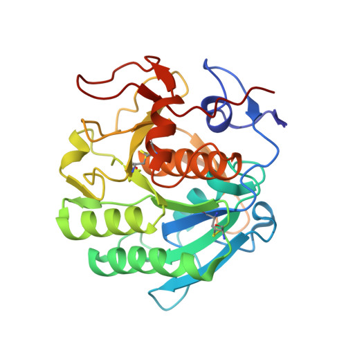
    In this work, a novel crystal growth method termed suspended drop crystallization has been developed. Unlike traditional methods, this technique involves mixing protein and precipitant directly on an electron microscopy grid without any additional support layers. The grid is then suspended within a crystallization chamber designed in-house, allowing for vapor diffusion to occur from both sides of the drop. A UV-transparent window above and below the grid enables the monitoring of crystal growth via light, UV or fluorescence microscopy. Once crystals have formed, the grid can be removed and utilized for X-ray crystallography or microcrystal electron diffraction (MicroED) directly without having to manipulate the crystals. To demonstrate the efficacy of this method, crystals of the enzyme proteinase K were grown and its structure was determined by MicroED following focused ion beam/scanning electron microscopy milling to render the sample thin enough for cryoEM. Suspended drop crystallization overcomes many of the challenges associated with sample preparation, providing an alternative workflow for crystals embedded in viscous media, sensitive to mechanical stress and/or subject to preferred orientation on electron microscopy grids.


  • Organizational Affiliation

    Departments of Biological Chemistry and Physiology, University of California, Los Angeles, CA, USA.


Macromolecules
Find similar proteins by:  (by identity cutoff)  |  3D Structure
Entity ID: 1
MoleculeChains Sequence LengthOrganismDetailsImage
Proteinase K279Parengyodontium albumMutation(s): 0 
EC: 3.4.21.64
UniProt
Find proteins for P06873 (Parengyodontium album)
Explore P06873 
Go to UniProtKB:  P06873
Entity Groups  
Sequence Clusters30% Identity50% Identity70% Identity90% Identity95% Identity100% Identity
UniProt GroupP06873
Sequence Annotations
Expand
  • Reference Sequence
Experimental Data & Validation

Experimental Data

  • Method: ELECTRON CRYSTALLOGRAPHY
  • Resolution: 2.10 Å
  • R-Value Free: 0.294 
  • R-Value Work: 0.255 
  • R-Value Observed: 0.259 
  • Space Group: P 43 21 2
Unit Cell:
Length ( Å )Angle ( ˚ )
a = 68.26α = 90
b = 68.26β = 90
c = 101.95γ = 90
Software Package:
Software NamePurpose
PHENIXrefinement
EM Software:
TaskSoftware PackageVersion
MODEL REFINEMENTPHENIX

Structure Validation

View Full Validation Report


Entry History & Funding Information

Deposition Data


Funding OrganizationLocationGrant Number
National Institutes of Health/National Institute of General Medical Sciences (NIH/NIGMS)United StatesP41GM136508
Department of Defense (DOD, United States)United StatesHDTRA1-21-1-0004
Howard Hughes Medical Institute (HHMI)United States--

Revision History  (Full details and data files)

  • Version 1.0: 2023-05-31
    Type: Initial release
  • Version 1.1: 2023-06-14
    Changes: Database references
  • Version 1.2: 2023-07-12
    Changes: Database references