8SD7

Carbonic anhydrase II radiation damage RT 61-90


Experimental Data Snapshot

  • Method: X-RAY DIFFRACTION
  • Resolution: 1.70 Å
  • R-Value Free: 0.153 
  • R-Value Work: 0.132 
  • R-Value Observed: 0.133 

wwPDB Validation   3D Report Full Report


This is version 1.0 of the entry. See complete history


Literature

XFEL structure of carbonic anhydrase II: a comparative study of XFEL, NMR, X-ray and neutron structures.

Hull, J.A.Lee, C.Kim, J.K.Lim, S.W.Park, J.Park, S.Lee, S.J.Park, G.Eom, I.Kim, M.Hyun, H.Combs, J.E.Andring, J.T.Lomelino, C.Kim, C.U.McKenna, R.

(2024) Acta Crystallogr D Struct Biol 80: 194-202

  • DOI: https://doi.org/10.1107/S2059798324000482
  • Primary Citation of Related Structures:  
    8SD1, 8SD6, 8SD7, 8SD8, 8SD9, 8SF1

  • PubMed Abstract: 

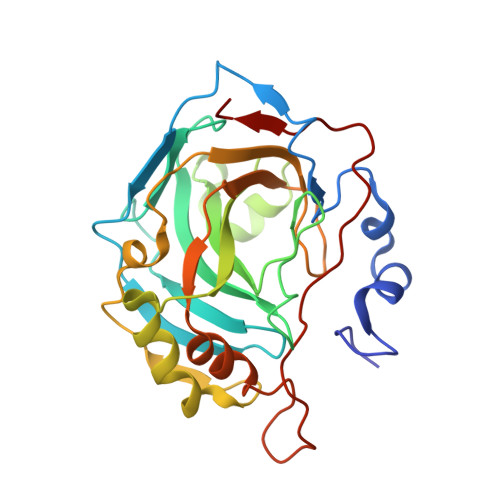
    The combination of X-ray free-electron lasers (XFELs) with serial femtosecond crystallography represents cutting-edge technology in structural biology, allowing the study of enzyme reactions and dynamics in real time through the generation of `molecular movies'. This technology combines short and precise high-energy X-ray exposure to a stream of protein microcrystals. Here, the XFEL structure of carbonic anhydrase II, a ubiquitous enzyme responsible for the interconversion of CO 2 and bicarbonate, is reported, and is compared with previously reported NMR and synchrotron X-ray and neutron single-crystal structures.


  • Organizational Affiliation

    Department of Biochemistry and Molecular Biology, College of Medicine, University of Florida, Gainesville, FL 32610, USA.


Macromolecules
Find similar proteins by:  (by identity cutoff)  |  3D Structure
Entity ID: 1
MoleculeChains Sequence LengthOrganismDetailsImage
Carbonic anhydrase 2260Homo sapiensMutation(s): 0 
Gene Names: CA2
EC: 4.2.1.1
UniProt & NIH Common Fund Data Resources
Find proteins for P00918 (Homo sapiens)
Explore P00918 
Go to UniProtKB:  P00918
PHAROS:  P00918
GTEx:  ENSG00000104267 
Entity Groups  
Sequence Clusters30% Identity50% Identity70% Identity90% Identity95% Identity100% Identity
UniProt GroupP00918
Sequence Annotations
Expand
  • Reference Sequence
Small Molecules
Ligands 1 Unique
IDChains Name / Formula / InChI Key2D Diagram3D Interactions
ZN
Query on ZN

Download Ideal Coordinates CCD File 
B [auth A]ZINC ION
Zn
PTFCDOFLOPIGGS-UHFFFAOYSA-N
Experimental Data & Validation

Experimental Data

  • Method: X-RAY DIFFRACTION
  • Resolution: 1.70 Å
  • R-Value Free: 0.153 
  • R-Value Work: 0.132 
  • R-Value Observed: 0.133 
  • Space Group: P 1 21 1
Unit Cell:
Length ( Å )Angle ( ˚ )
a = 42.713α = 90
b = 41.578β = 104.44
c = 72.707γ = 90
Software Package:
Software NamePurpose
PHENIXrefinement
Aimlessdata scaling
XDSdata reduction
PHENIXphasing

Structure Validation

View Full Validation Report


Entry History & Funding Information

Deposition Data


Funding OrganizationLocationGrant Number
Not funded--

Revision History  (Full details and data files)

  • Version 1.0: 2024-03-13
    Type: Initial release