8SBM | pdb_00008sbm

Crystal structure of the wild-type Catalytic ATP-binding domain of Mtb DosS


Experimental Data Snapshot

  • Method: X-RAY DIFFRACTION
  • Resolution: 1.47 Å
  • R-Value Free: 
    0.193 (Depositor), 0.193 (DCC) 
  • R-Value Work: 
    0.153 (Depositor), 0.153 (DCC) 
  • R-Value Observed: 
    0.155 (Depositor) 

Starting Model: in silico
View more details

wwPDB Validation 3D Report Full Report

Validation slider image for 8SBM

This is version 1.1 of the entry. See complete history

Literature

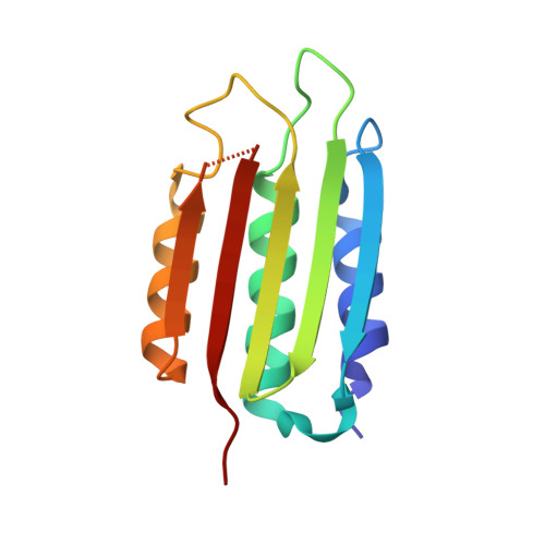
Understanding ATP Binding to DosS Catalytic Domain with a Short ATP-Lid.

Larson, G.W.Windsor, P.K.Smithwick, E.Shi, K.Aihara, H.Rama Damodaran, A.Bhagi-Damodaran, A.

(2023) Biochemistry 62: 3283-3292

  • DOI: https://doi.org/10.1021/acs.biochem.3c00306
  • Primary Citation Related Structures: 
    8SBM

  • PubMed Abstract: 

    DosS is a heme-containing histidine kinase that triggers dormancy transformation in Mycobacterium tuberculosis . Sequence comparison of the catalytic ATP-binding (CA) domain of DosS to other well-studied histidine kinases reveals a short ATP-lid. This feature has been thought to block binding of ATP to DosS's CA domain in the absence of interactions with DosS's dimerization and histidine phospho-transfer (DHp) domain. Here, we use a combination of computational modeling, structural biology, and biophysical studies to re-examine ATP-binding modalities in DosS. We show that the closed-lid conformation observed in crystal structures of DosS CA is caused by the presence of Zn 2+ in the ATP binding pocket that coordinates with Glu537 on the ATP-lid. Furthermore, circular dichroism studies and comparisons of DosS CA's crystal structure with its AlphaFold model and homologous DesK reveal that residues 503-507 that appear as a random coil in the Zn 2+ -coordinated crystal structure are in fact part of the N-box α helix needed for efficient ATP binding. Such random-coil transformation of an N-box α helix turn and the closed-lid conformation are both artifacts arising from large millimolar Zn 2+ concentrations used in DosS CA crystallization buffers. In contrast, in the absence of Zn 2+ , the short ATP-lid of DosS CA has significant conformational flexibility and can effectively bind AMP-PNP ( K d = 53 ± 13 μM), a non-hydrolyzable ATP analog. Furthermore, the nucleotide affinity remains unchanged when CA is conjugated to the DHp domain ( K d = 51 ± 6 μM). In all, our findings reveal that the short ATP-lid of DosS CA does not hinder ATP binding and provide insights that extend to 2988 homologous bacterial proteins containing such ATP-lids.


  • Organizational Affiliation
    • Department of Chemistry, University of Minnesota, Minneapolis, Minnesota 55455, United States.

Macromolecule Content 

  • Total Structure Weight: 27.25 kDa 
  • Atom Count: 2,170 
  • Modeled Residue Count: 247 
  • Deposited Residue Count: 252 
  • Unique protein chains: 1

Macromolecules

Find similar proteins by:|  3D Structure
Entity ID: 1
MoleculeChains  Sequence LengthOrganismDetailsImage
GAF domain-containing protein
A, B
126Mycobacterium tuberculosisMutation(s): 0 
Gene Names: GJE03_16455
EC: 2.7.13.3
UniProt
Find proteins for P9WGK3 (Mycobacterium tuberculosis (strain ATCC 25618 / H37Rv))
Explore P9WGK3 
Go to UniProtKB:  P9WGK3
Entity Groups
Sequence Clusters30% Identity50% Identity70% Identity90% Identity95% Identity100% Identity
UniProt GroupP9WGK3
Sequence Annotations
Expand
Reference Sequence

Small Molecules

Ligands 3 Unique
IDChains Name / Formula / InChI Key2D Diagram3D Interactions
ZN

Query on ZN


Download:Ideal Coordinates CCD File
C [auth A],
H [auth A],
J [auth B],
N [auth B]
ZINC ION
Zn
PTFCDOFLOPIGGS-UHFFFAOYSA-N
EDO

Query on EDO


Download:Ideal Coordinates CCD File
D [auth A]
E [auth A]
F [auth A]
G [auth A]
I [auth A]
D [auth A],
E [auth A],
F [auth A],
G [auth A],
I [auth A],
K [auth B],
L [auth B],
M [auth B]
1,2-ETHANEDIOL
C2 H6 O2
LYCAIKOWRPUZTN-UHFFFAOYSA-N
NA

Query on NA


Download:Ideal Coordinates CCD File
O [auth B]SODIUM ION
Na
FKNQFGJONOIPTF-UHFFFAOYSA-N

Experimental Data & Validation

Experimental Data

  • Method: X-RAY DIFFRACTION
  • Resolution: 1.47 Å
  • R-Value Free:  0.193 (Depositor), 0.193 (DCC) 
  • R-Value Work:  0.153 (Depositor), 0.153 (DCC) 
  • R-Value Observed: 0.155 (Depositor) 
Space Group: C 1 2 1
Unit Cell:
Length ( Å )Angle ( ˚ )
a = 54.438α = 90
b = 61.775β = 107.54
c = 72.234γ = 90
Software Package:
Software NamePurpose
PHENIXrefinement
Aimlessdata scaling
PDB_EXTRACTdata extraction
XDSdata reduction
PHASERphasing

Structure Validation

View Full Validation Report


Entry History 

& Funding Information

Deposition Data


Funding OrganizationLocationGrant Number
National Institutes of Health/National Institute of General Medical Sciences (NIH/NIGMS)United StatesR35-GM118047

Revision History  (Full details and data files)

  • Version 1.0: 2023-11-08
    Type: Initial release
  • Version 1.1: 2023-11-29
    Changes: Database references