8RXA | pdb_00008rxa

ACDC domain of AP2-O5 from Plasmodium falciparum


Experimental Data Snapshot

  • Method: X-RAY DIFFRACTION
  • Resolution: 1.75 Å
  • R-Value Free: 
    0.240 (Depositor), 0.255 (DCC) 
  • R-Value Work: 
    0.208 (Depositor) 
  • R-Value Observed: 
    0.210 (Depositor) 

Starting Model: in silico
View more details

wwPDB Validation 3D Report Full Report

Validation slider image for 8RXA

This is version 2.2 of the entry. See complete history

Literature

Structural characterization of the ACDC domain from ApiAP2 proteins, a potential molecular target against apicomplexan parasites.

Le Berre, M.Tubiana, T.Reutersward Waldner, P.Lazar, N.Li de la Sierra-Gallay, I.Santos, J.M.Llinas, M.Nessler, S.

(2025) Acta Crystallogr D Struct Biol 81: 38-48

  • DOI: https://doi.org/10.1107/S2059798324012518
  • Primary Citation Related Structures: 
    8RWU, 8RXA, 8RXO

  • PubMed Abstract: 

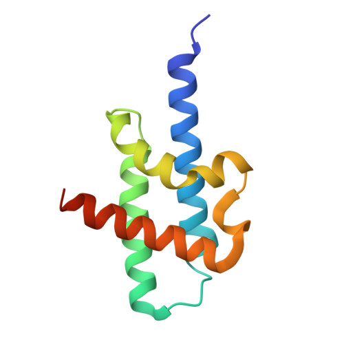
    The apicomplexan AP2 (ApiAP2) proteins are the best characterized family of DNA-binding proteins in Plasmodium spp. malaria parasites. Apart from the AP2 DNA-binding domain, there is little sequence similarity between ApiAP2 proteins. However, a conserved AP2-coincident domain mostly at the C-terminus (ACDC domain) is observed in a subset of the ApiAP2 proteins. The structure and function of this domain remain unknown. We report two crystal structures of ACDC domains derived from distinct Plasmodium ApiAP2 proteins, revealing a conserved, unique, noncanonical, four-helix bundle architecture. We used these structures to perform in silico docking calculations against a library of known antimalarial compounds and identified potential small-molecule ligands that bind in a highly conserved hydrophobic pocket that is present in all apicomplexan ACDC domains. These ligands provide a new molecular basis for the future design of ACDC inhibitors.


  • Organizational Affiliation
    • Institute for Integrative Biology of the Cell (I2BC), Université Paris-Saclay, CEA, CNRS, 91198 Gif-sur-Yvette, France.

Macromolecule Content 

  • Total Structure Weight: 26.14 kDa 
  • Atom Count: 1,704 
  • Modeled Residue Count: 197 
  • Deposited Residue Count: 218 
  • Unique protein chains: 1

Macromolecules

Find similar proteins by:|  3D Structure
Entity ID: 1
MoleculeChains  Sequence LengthOrganismDetailsImage
AP2 domain transcription factor AP2-O5, putative
A, B
109Plasmodium falciparum 3D7Mutation(s): 0 
Gene Names: PF3D7_1449500
UniProt
Find proteins for Q8IKY0 (Plasmodium falciparum (isolate 3D7))
Explore Q8IKY0 
Go to UniProtKB:  Q8IKY0
Entity Groups
Sequence Clusters30% Identity50% Identity70% Identity90% Identity95% Identity100% Identity
UniProt GroupQ8IKY0
Sequence Annotations
Expand
Reference Sequence

Experimental Data & Validation

Experimental Data

  • Method: X-RAY DIFFRACTION
  • Resolution: 1.75 Å
  • R-Value Free:  0.240 (Depositor), 0.255 (DCC) 
  • R-Value Work:  0.208 (Depositor) 
  • R-Value Observed: 0.210 (Depositor) 
Space Group: P 21 21 2
Unit Cell:
Length ( Å )Angle ( ˚ )
a = 58.93α = 90
b = 108.93β = 90
c = 34.01γ = 90
Software Package:
Software NamePurpose
PHENIXrefinement
XDSdata scaling
XDSdata reduction
PHASERphasing

Structure Validation

View Full Validation Report


Entry History 

& Funding Information

Deposition Data


Funding OrganizationLocationGrant Number
Not funded--

Revision History  (Full details and data files)

  • Version 1.0: 2024-03-06
    Type: Initial release
  • Version 2.0: 2024-12-25
    Type: Coordinate replacement
    Reason: Model completeness
    Changes: Advisory, Atomic model, Data collection, Database references, Derived calculations, Polymer sequence, Refinement description, Source and taxonomy, Structure summary
  • Version 2.1: 2025-01-22
    Changes: Data collection, Database references, Structure summary
  • Version 2.2: 2025-01-29
    Changes: Database references