8RI3 | pdb_00008ri3

Crystal structure of transplatin/B-DNA adduct obtained upon 7 days of soaking


Experimental Data Snapshot

  • Method: X-RAY DIFFRACTION
  • Resolution: 1.40 Å
  • R-Value Free: 
    0.257 (Depositor), 0.262 (DCC) 
  • R-Value Work: 
    0.216 (Depositor), 0.222 (DCC) 

Starting Model: experimental
View more details

wwPDB Validation 3D Report Full Report

Validation slider image for 8RI3

This is version 1.0 of the entry. See complete history

Literature

On the mechanism of action of arsenoplatins: arsenoplatin-1 binding to a B-DNA dodecamer.

Troisi, R.Tito, G.Ferraro, G.Sica, F.Massai, L.Geri, A.Cirri, D.Messori, L.Merlino, A.

(2024) Dalton Trans 53: 3476-3483

  • DOI: https://doi.org/10.1039/d3dt04302a
  • Primary Citation Related Structures: 
    8C62, 8C63, 8C64, 8RI3, 8RI5

  • PubMed Abstract: 

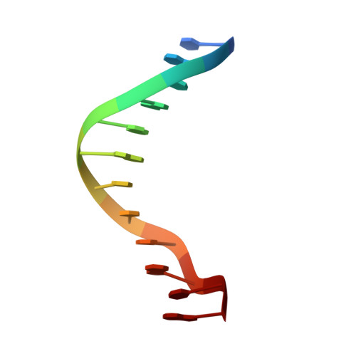
    The reaction of Pt-based anticancer agents with arsenic trioxide affords robust complexes known as arsenoplatins. The prototype of this family of anticancer compounds is arsenoplatin-1 (AP-1) that contains an As(OH) 2 fragment linked to a Pt(II) moiety derived from cisplatin. Crystallographic and spectrometric studies of AP-1 binding to a B-DNA double helix dodecamer are presented here, in comparison with cisplatin and transplatin. Results reveal that AP-1, cisplatin and transplatin react differently with the DNA model system. Notably, in the AP-1/DNA systems, the Pt-As bond can break down with time and As-containing fragments can be released. These results have implications for the understanding of the mechanism of action of arsenoplatins.


  • Organizational Affiliation
    • Department of Chemical Sciences, University of Naples Federico II, Complesso Universitario di Monte Sant'Angelo, via Cintia, 80126, Naples, Italy. antonello.merlino@unina.it.

Macromolecule Content 

  • Total Structure Weight: 8.02 kDa 
  • Atom Count: 603 
  • Modeled Residue Count: 24 
  • Deposited Residue Count: 24 
  • Unique nucleic acid chains: 1

Macromolecules

Find similar nucleic acids by:  Sequence
Entity ID: 1
MoleculeChains LengthOrganismImage
DNA (5'-D(*CP*GP*CP*GP*AP*AP*TP*TP*CP*GP*CP*G)-3')
A, B
12synthetic construct
Sequence Annotations
Expand
Reference Sequence

Small Molecules

Ligands 4 Unique
IDChains Name / Formula / InChI Key2D Diagram3D Interactions
PT
(Subject of Investigation/LOI)

Query on PT


Download:Ideal Coordinates CCD File
D [auth A],
F [auth A],
I [auth B]
PLATINUM (II) ION
Pt
HRGDZIGMBDGFTC-UHFFFAOYSA-N
CL

Query on CL


Download:Ideal Coordinates CCD File
H [auth A]CHLORIDE ION
Cl
VEXZGXHMUGYJMC-UHFFFAOYSA-M
MG

Query on MG


Download:Ideal Coordinates CCD File
C [auth A]MAGNESIUM ION
Mg
JLVVSXFLKOJNIY-UHFFFAOYSA-N
NH3

Query on NH3


Download:Ideal Coordinates CCD File
E [auth A],
G [auth A],
J [auth B]
AMMONIA
H3 N
QGZKDVFQNNGYKY-UHFFFAOYSA-N

Experimental Data & Validation

Experimental Data

  • Method: X-RAY DIFFRACTION
  • Resolution: 1.40 Å
  • R-Value Free:  0.257 (Depositor), 0.262 (DCC) 
  • R-Value Work:  0.216 (Depositor), 0.222 (DCC) 
Space Group: P 21 21 21
Unit Cell:
Length ( Å )Angle ( ˚ )
a = 25.56α = 90
b = 40.39β = 90
c = 65.67γ = 90
Software Package:
Software NamePurpose
REFMACrefinement
autoPROCdata reduction
autoPROCdata scaling
PHASERphasing

Structure Validation

View Full Validation Report


Entry History 

& Funding Information

Deposition Data


Funding OrganizationLocationGrant Number
Not funded--

Revision History  (Full details and data files)

  • Version 1.0: 2024-02-28
    Type: Initial release