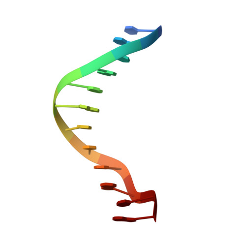
8RI3

Crystal structure of transplatin/B-DNA adduct obtained upon 7 days of soaking


Experimental Data Snapshot

  • Method: X-RAY DIFFRACTION
  • Resolution: 1.40 Å
  • R-Value Free: 0.257 
  • R-Value Work: 0.216 

wwPDB Validation   3D Report Full Report


This is version 1.0 of the entry. See complete history


Literature

On the mechanism of action of arsenoplatins: arsenoplatin-1 binding to a B-DNA dodecamer.

Troisi, R.Tito, G.Ferraro, G.Sica, F.Massai, L.Geri, A.Cirri, D.Messori, L.Merlino, A.

(2024) Dalton Trans 53: 3476-3483

  • DOI: https://doi.org/10.1039/d3dt04302a
  • Primary Citation of Related Structures:  
    8C62, 8C63, 8C64, 8RI3, 8RI5

  • PubMed Abstract: 

    The reaction of Pt-based anticancer agents with arsenic trioxide affords robust complexes known as arsenoplatins. The prototype of this family of anticancer compounds is arsenoplatin-1 (AP-1) that contains an As(OH) 2 fragment linked to a Pt(II) moiety derived from cisplatin. Crystallographic and spectrometric studies of AP-1 binding to a B-DNA double helix dodecamer are presented here, in comparison with cisplatin and transplatin. Results reveal that AP-1, cisplatin and transplatin react differently with the DNA model system. Notably, in the AP-1/DNA systems, the Pt-As bond can break down with time and As-containing fragments can be released. These results have implications for the understanding of the mechanism of action of arsenoplatins.


  • Organizational Affiliation

    Department of Chemical Sciences, University of Naples Federico II, Complesso Universitario di Monte Sant'Angelo, via Cintia, 80126, Naples, Italy. antonello.merlino@unina.it.


Macromolecules

Find similar nucleic acids by:  Sequence   |   3D Structure  

Entity ID: 1
MoleculeChains LengthOrganismImage
DNA (5'-D(*CP*GP*CP*GP*AP*AP*TP*TP*CP*GP*CP*G)-3')
A, B
12synthetic construct
Sequence Annotations
Expand
  • Reference Sequence
Small Molecules
Ligands 4 Unique
IDChains Name / Formula / InChI Key2D Diagram3D Interactions
PT (Subject of Investigation/LOI)
Query on PT

Download Ideal Coordinates CCD File 
D [auth A],
F [auth A],
I [auth B]
PLATINUM (II) ION
Pt
HRGDZIGMBDGFTC-UHFFFAOYSA-N
CL
Query on CL

Download Ideal Coordinates CCD File 
H [auth A]CHLORIDE ION
Cl
VEXZGXHMUGYJMC-UHFFFAOYSA-M
MG
Query on MG

Download Ideal Coordinates CCD File 
C [auth A]MAGNESIUM ION
Mg
JLVVSXFLKOJNIY-UHFFFAOYSA-N
NH3
Query on NH3

Download Ideal Coordinates CCD File 
E [auth A],
G [auth A],
J [auth B]
AMMONIA
H3 N
QGZKDVFQNNGYKY-UHFFFAOYSA-N
Experimental Data & Validation

Experimental Data

  • Method: X-RAY DIFFRACTION
  • Resolution: 1.40 Å
  • R-Value Free: 0.257 
  • R-Value Work: 0.216 
  • Space Group: P 21 21 21
Unit Cell:
Length ( Å )Angle ( ˚ )
a = 25.56α = 90
b = 40.39β = 90
c = 65.67γ = 90
Software Package:
Software NamePurpose
REFMACrefinement
autoPROCdata reduction
autoPROCdata scaling
PHASERphasing

Structure Validation

View Full Validation Report


Entry History & Funding Information

Deposition Data


Funding OrganizationLocationGrant Number
Not funded--

Revision History  (Full details and data files)

  • Version 1.0: 2024-02-28
    Type: Initial release