


Ruthenium Polypyridyl complex intercalating in Major and Minor Groove DNA
Abdullrahman, A., McQuaid, K., Cardin, C.J., Hall, J.P.To be published.
Experimental Data Snapshot
Entity ID: 1 | ||||
| Molecule | Chains | Length | Organism | Image |
|---|---|---|---|---|
| DNA (5'-D(*GP*AP*AP*TP*AP*GP*G)-3') | 7 | synthetic construct | 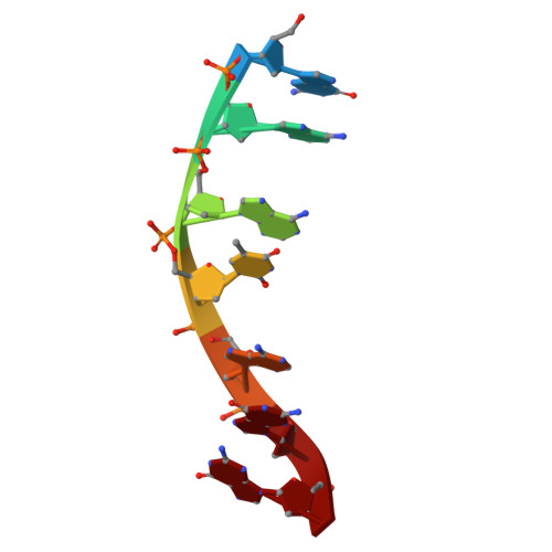 | |
Sequence AnnotationsExpand | ||||
Reference Sequence | ||||
Entity ID: 2 | ||||
| Molecule | Chains | Length | Organism | Image |
|---|---|---|---|---|
| DNA (5'-D(*CP*CP*TP*AP*TP*TP*C)-3') | 7 | synthetic construct |  | |
Sequence AnnotationsExpand | ||||
Reference Sequence | ||||
| Ligands 3 Unique | |||||
|---|---|---|---|---|---|
| ID | Chains | Name / Formula / InChI Key | 2D Diagram | 3D Interactions | |
| KSB (Subject of Investigation/LOI) Download:Ideal Coordinates CCD File | E [auth A], J [auth B], O [auth C], T [auth D] | lambda-[Ru(tap2-dppz-CN)]2+ C39 H21 N13 Ru HNFPSJMHTKCUIN-UHFFFAOYSA-N |  | ||
| CL Download:Ideal Coordinates CCD File | H [auth A] I [auth A] M [auth B] N [auth B] S [auth C] | CHLORIDE ION Cl VEXZGXHMUGYJMC-UHFFFAOYSA-M |  | ||
| MG Download:Ideal Coordinates CCD File | F [auth A] G [auth A] K [auth B] L [auth B] P [auth C] | MAGNESIUM ION Mg JLVVSXFLKOJNIY-UHFFFAOYSA-N |  | ||
| Length ( Å ) | Angle ( ˚ ) |
|---|---|
| a = 32.676 | α = 90 |
| b = 32.676 | β = 90 |
| c = 103.988 | γ = 90 |
| Software Name | Purpose |
|---|---|
| PHENIX | refinement |
| DIALS | data reduction |
| xia2 | data scaling |
| SHELXDE | phasing |
| Funding Organization | Location | Grant Number |
|---|---|---|
| H2020 Marie Curie Actions of the European Commission | European Union | H2020-MSCA-INT-2019-861 |


