8R0J

Crystal structure of the retromer complex VPS29/VPS35 with the ligand bis-1,3-phenyl guanylhydrazone, 2a


Experimental Data Snapshot

  • Method: X-RAY DIFFRACTION
  • Resolution: 2.40 Å
  • R-Value Free: 0.250 
  • R-Value Work: 0.185 

wwPDB Validation   3D Report Full Report


Ligand Structure Quality Assessment 


This is version 1.0 of the entry. See complete history


Literature

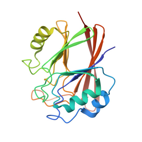
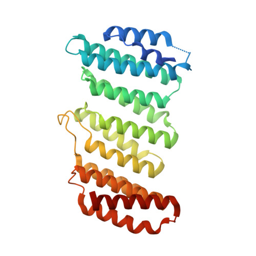
Stabilization of the retromer complex: Analysis of novel binding sites of bis-1,3-phenyl guanylhydrazone 2a to the VPS29/VPS35 interface.

Fagnani, E.Boni, F.Seneci, P.Gornati, D.Muzio, L.Mastrangelo, E.Milani, M.

(2024) Comput Struct Biotechnol J 23: 1088-1093

  • DOI: https://doi.org/10.1016/j.csbj.2024.02.026
  • Primary Citation of Related Structures:  
    8R02, 8R0J

  • PubMed Abstract: 

    The stabilization of the retromer protein complex can be effective in the treatment of different neurological disorders. Following the identification of bis-1,3-phenyl guanylhydrazone 2a as an effective new compound for the treatment of amyotrophic lateral sclerosis, in this work we analyze the possible binding sites of this molecule to the VPS35/VPS29 dimer of the retromer complex. Our results show that the affinity for different sites of the protein assembly depends on compound charge and therefore slight changes in the cell microenvironment could promote different binding states. Finally, we describe a novel binding site located in a deep cleft between VPS29 and VPS35 that should be further explored to select novel molecular chaperones for the stabilization of the retromer complex.


  • Organizational Affiliation

    Biophysics Institute, CNR-IBF, Via Corti 12, I-20133 Milano, Italy.


Macromolecules
Find similar proteins by:  (by identity cutoff)  |  3D Structure
Entity ID: 1
MoleculeChains Sequence LengthOrganismDetailsImage
Vacuolar protein sorting-associated protein 29
A, B
185Homo sapiensMutation(s): 0 
Gene Names: VPS29
UniProt & NIH Common Fund Data Resources
Find proteins for Q9UBQ0 (Homo sapiens)
Explore Q9UBQ0 
Go to UniProtKB:  Q9UBQ0
PHAROS:  Q9UBQ0
GTEx:  ENSG00000111237 
Entity Groups  
Sequence Clusters30% Identity50% Identity70% Identity90% Identity95% Identity100% Identity
UniProt GroupQ9UBQ0
Sequence Annotations
Expand
  • Reference Sequence
Find similar proteins by:  (by identity cutoff)  |  3D Structure
Entity ID: 2
MoleculeChains Sequence LengthOrganismDetailsImage
Vacuolar protein sorting-associated protein 35
C, D
306Homo sapiensMutation(s): 0 
Gene Names: VPS35
UniProt & NIH Common Fund Data Resources
Find proteins for Q96QK1 (Homo sapiens)
Explore Q96QK1 
Go to UniProtKB:  Q96QK1
PHAROS:  Q96QK1
GTEx:  ENSG00000069329 
Entity Groups  
Sequence Clusters30% Identity50% Identity70% Identity90% Identity95% Identity100% Identity
UniProt GroupQ96QK1
Sequence Annotations
Expand
  • Reference Sequence
Small Molecules
Ligands 2 Unique
IDChains Name / Formula / InChI Key2D Diagram3D Interactions
XFZ (Subject of Investigation/LOI)
Query on XFZ

Download Ideal Coordinates CCD File 
E [auth C]Bis-1,3-phenyl guanylhydrazon
C10 H14 N8
XMHHBYPIBVIERY-IAGONARPSA-N
TRS
Query on TRS

Download Ideal Coordinates CCD File 
F [auth D]2-AMINO-2-HYDROXYMETHYL-PROPANE-1,3-DIOL
C4 H12 N O3
LENZDBCJOHFCAS-UHFFFAOYSA-O
Experimental Data & Validation

Experimental Data

  • Method: X-RAY DIFFRACTION
  • Resolution: 2.40 Å
  • R-Value Free: 0.250 
  • R-Value Work: 0.185 
  • Space Group: P 21 21 21
Unit Cell:
Length ( Å )Angle ( ˚ )
a = 48.15α = 90
b = 130.34β = 90
c = 146.89γ = 90
Software Package:
Software NamePurpose
REFMACrefinement
XDSdata reduction
XSCALEdata scaling
MOLREPphasing

Structure Validation

View Full Validation Report


Ligand Structure Quality Assessment 


Entry History & Funding Information

Deposition Data


Funding OrganizationLocationGrant Number
Other privateItalyTRAILER

Revision History  (Full details and data files)

  • Version 1.0: 2024-03-27
    Type: Initial release