8QV0

Structure of the native microtubule lattice nucleated from the yeast spindle pole body


Experimental Data Snapshot

  • Method: ELECTRON MICROSCOPY
  • Resolution: 6.60 Å
  • Aggregation State: PARTICLE 
  • Reconstruction Method: SUBTOMOGRAM AVERAGING 

wwPDB Validation   3D Report Full Report


This is version 1.0 of the entry. See complete history


Literature

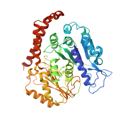
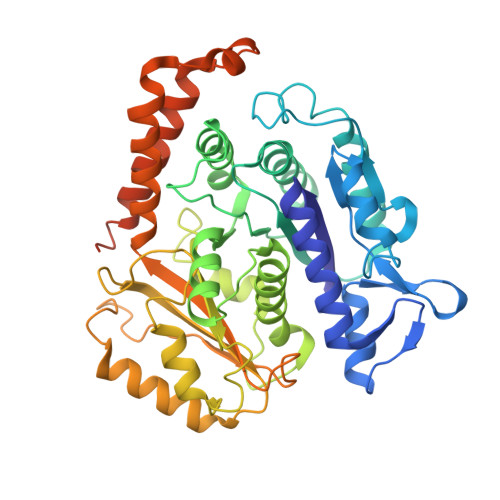
Structure of the native gamma-tubulin ring complex capping spindle microtubules.

Dendooven, T.Yatskevich, S.Burt, A.Chen, Z.A.Bellini, D.Rappsilber, J.Kilmartin, J.V.Barford, D.

(2024) Nat Struct Mol Biol 

  • DOI: https://doi.org/10.1038/s41594-024-01281-y
  • Primary Citation of Related Structures:  
    8QV0, 8QV2, 8QV3

  • PubMed Abstract: 

    Microtubule (MT) filaments, composed of α/β-tubulin dimers, are fundamental to cellular architecture, function and organismal development. They are nucleated from MT organizing centers by the evolutionarily conserved γ-tubulin ring complex (γTuRC). However, the molecular mechanism of nucleation remains elusive. Here we used cryo-electron tomography to determine the structure of the native γTuRC capping the minus end of a MT in the context of enriched budding yeast spindles. In our structure, γTuRC presents a ring of γ-tubulin subunits to seed nucleation of exclusively 13-protofilament MTs, adopting an active closed conformation to function as a perfect geometric template for MT nucleation. Our cryo-electron tomography reconstruction revealed that a coiled-coil protein staples the first row of α/β-tubulin of the MT to alternating positions along the γ-tubulin ring of γTuRC. This positioning of α/β-tubulin onto γTuRC suggests a role for the coiled-coil protein in augmenting γTuRC-mediated MT nucleation. Based on our results, we describe a molecular model for budding yeast γTuRC activation and MT nucleation.


  • Organizational Affiliation

    MRC Laboratory of Molecular Biology, Cambridge, UK. tdendooven@mrc-lmb.cam.ac.uk.


Macromolecules
Find similar proteins by:  (by identity cutoff)  |  3D Structure
Entity ID: 1
MoleculeChains Sequence LengthOrganismDetailsImage
Tubulin alpha-1 chain
A, C, D, E, F
A, C, D, E, F, G, H, I, J, K, L, M, N
447Saccharomyces cerevisiaeMutation(s): 0 
UniProt
Find proteins for P09733 (Saccharomyces cerevisiae (strain ATCC 204508 / S288c))
Explore P09733 
Go to UniProtKB:  P09733
Entity Groups  
Sequence Clusters30% Identity50% Identity70% Identity90% Identity95% Identity100% Identity
UniProt GroupP09733
Sequence Annotations
Expand
  • Reference Sequence
Find similar proteins by:  (by identity cutoff)  |  3D Structure
Entity ID: 2
MoleculeChains Sequence LengthOrganismDetailsImage
Tubulin beta chain
B, O, P, Q, R
B, O, P, Q, R, S, T, U, V, W, X, Y, Z
457Saccharomyces cerevisiaeMutation(s): 0 
UniProt
Find proteins for A0A6A5PXT5 (Saccharomyces cerevisiae)
Explore A0A6A5PXT5 
Go to UniProtKB:  A0A6A5PXT5
Entity Groups  
Sequence Clusters30% Identity50% Identity70% Identity90% Identity95% Identity100% Identity
UniProt GroupA0A6A5PXT5
Sequence Annotations
Expand
  • Reference Sequence
Experimental Data & Validation

Experimental Data

  • Method: ELECTRON MICROSCOPY
  • Resolution: 6.60 Å
  • Aggregation State: PARTICLE 
  • Reconstruction Method: SUBTOMOGRAM AVERAGING 
EM Software:
TaskSoftware PackageVersion
MODEL REFINEMENTPHENIX1.17.1_3660:

Structure Validation

View Full Validation Report


Entry History & Funding Information

Deposition Data


Funding OrganizationLocationGrant Number
Cancer Research UKUnited Kingdom--
Boehringer Ingelheim Fonds (BIF)Germany--
Medical Research Council (MRC, United Kingdom)United Kingdom--

Revision History  (Full details and data files)

  • Version 1.0: 2024-04-24
    Type: Initial release