8PYR

Crystal structure of the dual T-loop phosphorylated Cdk7/CycH/Mat1 complex


Experimental Data Snapshot

  • Method: X-RAY DIFFRACTION
  • Resolution: 2.15 Å
  • R-Value Free: 0.241 
  • R-Value Work: 0.220 
  • R-Value Observed: 0.223 

wwPDB Validation   3D Report Full Report


This is version 1.0 of the entry. See complete history


Literature

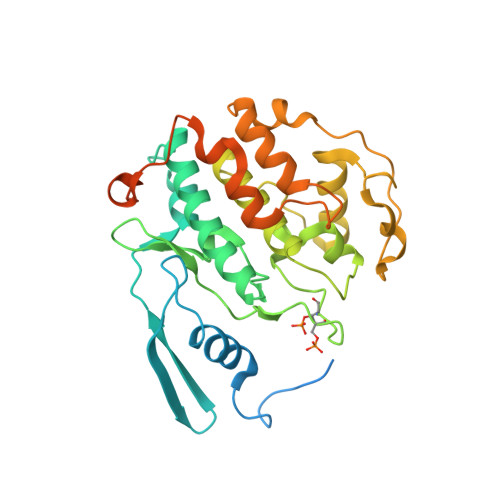
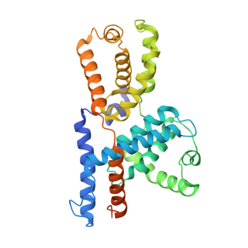
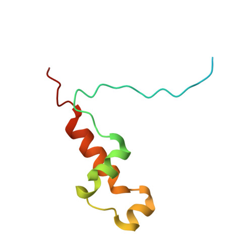
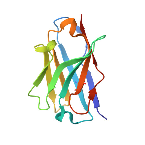
Structural basis of Cdk7 activation by dual T-loop phosphorylation.

Duster, R.Anand, K.Binder, S.C.Schmitz, M.Gatterdam, K.Fisher, R.P.Geyer, M.

(2024) Biorxiv 

  • DOI: https://doi.org/10.1101/2024.02.14.580246
  • Primary Citation of Related Structures:  
    8PYR

  • PubMed Abstract: 

    Cyclin-dependent kinase 7 (Cdk7) occupies a central position in cell-cycle and transcriptional regulation owing to its function as both a CDK-activating kinase (CAK) and part of the general transcription factor TFIIH. Cdk7 forms an active complex upon association with Cyclin H and Mat1, and its catalytic activity is regulated by two phosphorylations in the activation segment (T loop): the canonical activating modification at T170 and another at S164. Here we report the crystal structure of the fully activated human Cdk7/Cyclin H/Mat1 complex containing both T-loop phosphorylations. Whereas pT170 coordinates a set of basic residues conserved in other CDKs, pS164 nucleates an arginine network involving all three subunits that is unique to the ternary Cdk7 complex. We identify differential dependencies of kinase activity and substrate recognition on individual phosphorylations within the Cdk7 T loop. The CAK function of Cdk7 is not affected by T-loop phosphorylation, whereas activity towards non-CDK substrates is increased several-fold by phosphorylation at T170. Moreover, dual T-loop phosphorylation at both T170 and S164 stimulates multi-site phosphorylation of transcriptional substrates-the RNA polymerase II (RNAPII) carboxy-terminal domain (CTD) and the SPT5 carboxy-terminal repeat (CTR) region. In human cells, Cdk7-regulatory phosphorylation is a two-step process in which phosphorylation of S164 precedes, and may prime, T170 phosphorylation. Thus, dual T-loop phosphorylation can regulate Cdk7 through multiple mechanisms, with pS164 supporting tripartite complex formation and possibly influencing Cdk7 processivity, while the canonical pT170 enhances kinase activity towards critical substrates involved in transcription.


  • Organizational Affiliation

    Institute of Structural Biology, University of Bonn, Venusberg-Campus 1, 53127 Bonn, Germany.


Macromolecules
Find similar proteins by:  (by identity cutoff)  |  3D Structure
Entity ID: 1
MoleculeChains Sequence LengthOrganismDetailsImage
Cyclin-dependent kinase 7
A, E
346Homo sapiensMutation(s): 0 
Gene Names: CDK7CAKCAK1CDKN7MO15STK1
EC: 2.7.11.22 (PDB Primary Data), 2.7.11.23 (PDB Primary Data)
UniProt & NIH Common Fund Data Resources
Find proteins for P50613 (Homo sapiens)
Explore P50613 
Go to UniProtKB:  P50613
PHAROS:  P50613
GTEx:  ENSG00000134058 
Entity Groups  
Sequence Clusters30% Identity50% Identity70% Identity90% Identity95% Identity100% Identity
UniProt GroupP50613
Sequence Annotations
Expand
  • Reference Sequence
Find similar proteins by:  (by identity cutoff)  |  3D Structure
Entity ID: 2
MoleculeChains Sequence LengthOrganismDetailsImage
Cyclin-H
B, F
323Homo sapiensMutation(s): 0 
Gene Names: CCNH
UniProt & NIH Common Fund Data Resources
Find proteins for P51946 (Homo sapiens)
Explore P51946 
Go to UniProtKB:  P51946
PHAROS:  P51946
GTEx:  ENSG00000134480 
Entity Groups  
Sequence Clusters30% Identity50% Identity70% Identity90% Identity95% Identity100% Identity
UniProt GroupP51946
Sequence Annotations
Expand
  • Reference Sequence
Find similar proteins by:  (by identity cutoff)  |  3D Structure
Entity ID: 3
MoleculeChains Sequence LengthOrganismDetailsImage
CDK-activating kinase assembly factor MAT1
C, G
82Homo sapiensMutation(s): 0 
Gene Names: MNAT1CAP35MAT1RNF66
UniProt & NIH Common Fund Data Resources
Find proteins for P51948 (Homo sapiens)
Explore P51948 
Go to UniProtKB:  P51948
PHAROS:  P51948
GTEx:  ENSG00000020426 
Entity Groups  
Sequence Clusters30% Identity50% Identity70% Identity90% Identity95% Identity100% Identity
UniProt GroupP51948
Sequence Annotations
Expand
  • Reference Sequence
Find similar proteins by:  (by identity cutoff)  |  3D Structure
Entity ID: 4
MoleculeChains Sequence LengthOrganismDetailsImage
Nanobody (VHH-RD7-04)
D, H
115Lama glamaMutation(s): 0 
Entity Groups  
Sequence Clusters30% Identity50% Identity70% Identity90% Identity95% Identity100% Identity
Sequence Annotations
Expand
  • Reference Sequence
Experimental Data & Validation

Experimental Data

  • Method: X-RAY DIFFRACTION
  • Resolution: 2.15 Å
  • R-Value Free: 0.241 
  • R-Value Work: 0.220 
  • R-Value Observed: 0.223 
  • Space Group: P 1 21 1
Unit Cell:
Length ( Å )Angle ( ˚ )
a = 119.86α = 90
b = 77.866β = 119.31
c = 121.872γ = 90
Software Package:
Software NamePurpose
PHENIXrefinement
XDSdata reduction
XSCALEdata scaling
PHASERphasing

Structure Validation

View Full Validation Report


Entry History & Funding Information

Deposition Data


Funding OrganizationLocationGrant Number
German Research Foundation (DFG)GermanyEXC2151-390873048

Revision History  (Full details and data files)

  • Version 1.0: 2024-03-06
    Type: Initial release