8PUQ

MetHemoglobin structure from serial synchrotron crystallography with fixed target


Experimental Data Snapshot

  • Method: X-RAY DIFFRACTION
  • Resolution: 1.95 Å
  • R-Value Free: 0.174 
  • R-Value Work: 0.125 

wwPDB Validation   3D Report Full Report


Ligand Structure Quality Assessment 


This is version 1.2 of the entry. See complete history


Literature

Anaerobic fixed-target serial crystallography using sandwiched silicon nitride membranes.

Bjelcic, M.Sigfridsson Clauss, K.G.V.Aurelius, O.Milas, M.Nan, J.Ursby, T.

(2023) Acta Crystallogr D Struct Biol 79: 1018-1025

  • DOI: https://doi.org/10.1107/S205979832300880X
  • Primary Citation of Related Structures:  
    8PUQ, 8PUR

  • PubMed Abstract: 

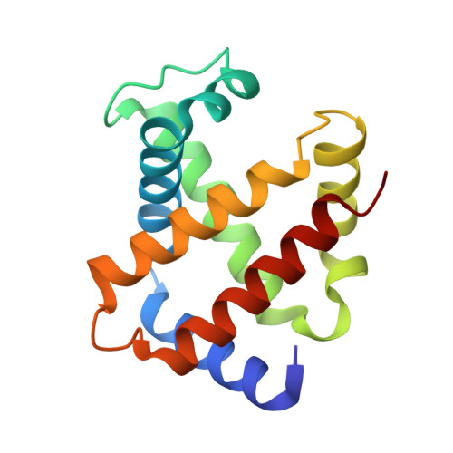
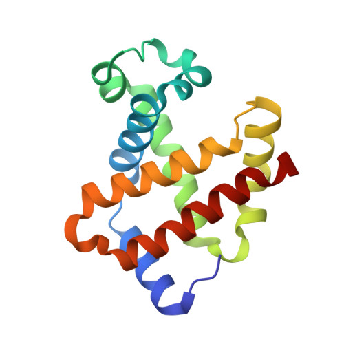
    In recent years, the emergence of serial crystallography, initially pioneered at X-ray free-electron lasers (XFELs), has sparked a growing interest in collecting macromolecular crystallographic data at room temperature. Various fixed-target serial crystallography techniques have been developed, ranging from commercially available chips to in-house designs implemented at different synchrotron facilities. Nevertheless, there is currently no commercially available chip (known to the authors) specifically designed for the direct handling of oxygen-sensitive samples. This study presents a methodology employing silicon nitride chips arranged in a `sandwich' configuration, enabling reliable room-temperature data collection from oxygen-sensitive samples. The method involves the utilization of a custom-made 3D-printed assembling tool and a MX sample holder. To validate the effectiveness of the proposed method, deoxyhemoglobin and methemoglobin samples were investigated using the BioMAX X-ray macromolecular crystallography beamline, the Balder X-ray absorption spectroscopy beamline and UV-Vis absorption spectroscopy.


  • Organizational Affiliation

    MAX IV Laboratory, Lund University, PO Box 118, SE-221 00 Lund, Sweden.


Macromolecules
Find similar proteins by:  (by identity cutoff)  |  3D Structure
Entity ID: 1
MoleculeChains Sequence LengthOrganismDetailsImage
Hemoglobin subunit alpha140Equus caballusMutation(s): 0 
UniProt
Find proteins for P01958 (Equus caballus)
Explore P01958 
Go to UniProtKB:  P01958
Entity Groups  
Sequence Clusters30% Identity50% Identity70% Identity90% Identity95% Identity100% Identity
UniProt GroupP01958
Sequence Annotations
Expand
  • Reference Sequence
Find similar proteins by:  (by identity cutoff)  |  3D Structure
Entity ID: 2
MoleculeChains Sequence LengthOrganismDetailsImage
Hemoglobin subunit beta146Equus caballusMutation(s): 0 
UniProt
Find proteins for P02062 (Equus caballus)
Explore P02062 
Go to UniProtKB:  P02062
Entity Groups  
Sequence Clusters30% Identity50% Identity70% Identity90% Identity95% Identity100% Identity
UniProt GroupP02062
Sequence Annotations
Expand
  • Reference Sequence
Small Molecules
Experimental Data & Validation

Experimental Data

  • Method: X-RAY DIFFRACTION
  • Resolution: 1.95 Å
  • R-Value Free: 0.174 
  • R-Value Work: 0.125 
  • Space Group: C 1 2 1
Unit Cell:
Length ( Å )Angle ( ˚ )
a = 108.53α = 90
b = 63.08β = 111.02
c = 54.69γ = 90
Software Package:
Software NamePurpose
REFMACrefinement
CrystFELdata reduction
CrystFELdata scaling
PHASERphasing

Structure Validation

View Full Validation Report


Ligand Structure Quality Assessment 


Entry History & Funding Information

Deposition Data


Funding OrganizationLocationGrant Number
Other government--

Revision History  (Full details and data files)

  • Version 1.0: 2023-10-18
    Type: Initial release
  • Version 1.1: 2023-11-01
    Changes: Database references
  • Version 1.2: 2023-11-08
    Changes: Database references