8PJN | pdb_00008pjn

Catalytic module of human CTLH E3 ligase bound to multiphosphorylated UBE2H~ubiquitin


Experimental Data Snapshot

  • Method: ELECTRON MICROSCOPY
  • Resolution: 3.40 Å
  • Aggregation State: PARTICLE 
  • Reconstruction Method: SINGLE PARTICLE 

wwPDB Validation 3D Report Full Report

Validation slider image for 8PJN

This is version 1.4 of the entry. See complete history. 

Literature

Multisite phosphorylation dictates selective E2-E3 pairing as revealed by Ubc8/UBE2H-GID/CTLH assemblies.

Chrustowicz, J., Sherpa, D., Li, J., Langlois, C.R., Papadopoulou, E.C., Vu, D.T., Hehl, L.A., Karayel, O., Beier, V., von Gronau, S., Muller, J., Prabu, J.R., Mann, M., Kleiger, G., Alpi, A.F., Schulman, B.A.

(2024) Mol Cell 84: 293

  • DOI: https://doi.org/10.1016/j.molcel.2023.11.027
  • Primary Citation Related Structures: 
    8PJN, 8PMQ

  • PubMed Abstract: 

    Ubiquitylation is catalyzed by coordinated actions of E3 and E2 enzymes. Molecular principles governing many important E3-E2 partnerships remain unknown, including those for RING-family GID/CTLH E3 ubiquitin ligases and their dedicated E2, Ubc8/UBE2H (yeast/human nomenclature). GID/CTLH-Ubc8/UBE2H-mediated ubiquitylation regulates biological processes ranging from yeast metabolic signaling to human development. Here, cryoelectron microscopy (cryo-EM), biochemistry, and cell biology reveal this exquisitely specific E3-E2 pairing through an unconventional catalytic assembly and auxiliary interactions 70-100 Å away, mediated by E2 multisite phosphorylation. Rather than dynamic polyelectrostatic interactions reported for other ubiquitylation complexes, multiple Ubc8/UBE2H phosphorylation sites within acidic CK2-targeted sequences specifically anchor the E2 C termini to E3 basic patches. Positions of phospho-dependent interactions relative to the catalytic domains correlate across evolution. Overall, our data show that phosphorylation-dependent multivalency establishes a specific E3-E2 partnership, is antagonistic with dephosphorylation, rigidifies the catalytic centers within a flexing GID E3-substrate assembly, and facilitates substrate collision with ubiquitylation active sites.


  • Organizational Affiliation: 
    • Department of Molecular Machines and Signaling, Max Planck Institute of Biochemistry, Martinsried 82152, Germany.

Macromolecule Content 

  • Total Structure Weight: 119.37 kDa 
  • Atom Count: 6,917 
  • Modeled Residue Count: 923 
  • Deposited Residue Count: 1,046 
  • Unique protein chains: 4

Macromolecules

Find similar proteins by:|  3D Structure
Entity ID: 1
MoleculeChains  Sequence LengthOrganismDetailsImage
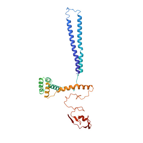
E3 ubiquitin-protein transferase RMND5AA [auth b]391Homo sapiensMutation(s): 0 
Gene Names: RMND5A
EC: 2.3.2.27
UniProt & NIH Common Fund Data Resources
Find proteins for Q9H871 (Homo sapiens)
Explore Q9H871 
Go to UniProtKB:  Q9H871
PHAROS:  Q9H871
GTEx:  ENSG00000153561 
Entity Groups
Sequence Clusters30% Identity50% Identity70% Identity90% Identity95% Identity100% Identity
UniProt GroupQ9H871
Sequence Annotations
Expand
Reference Sequence
Find similar proteins by:|  3D Structure
Entity ID: 2
MoleculeChains  Sequence LengthOrganismDetailsImage
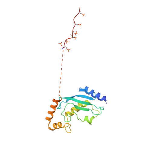
Ubiquitin-conjugating enzyme E2 HB [auth 2]183Homo sapiensMutation(s): 1 
Gene Names: UBE2H
EC: 2.3.2.23 (PDB Primary Data), 2.3.2.24 (PDB Primary Data)
UniProt & NIH Common Fund Data Resources
Find proteins for P62256 (Homo sapiens)
Explore P62256 
Go to UniProtKB:  P62256
PHAROS:  P62256
GTEx:  ENSG00000186591 
Entity Groups
Sequence Clusters30% Identity50% Identity70% Identity90% Identity95% Identity100% Identity
UniProt GroupP62256
Sequence Annotations
Expand
Reference Sequence
Find similar proteins by:|  3D Structure
Entity ID: 3
MoleculeChains  Sequence LengthOrganismDetailsImage
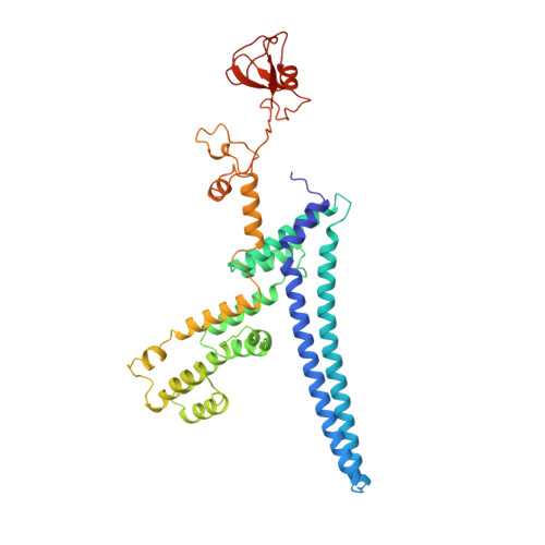
E3 ubiquitin-protein transferase MAEAC [auth i]396Homo sapiensMutation(s): 0 
Gene Names: MAEA, EMP, HLC10, PIG5
EC: 2.3.2.27
UniProt & NIH Common Fund Data Resources
Find proteins for Q7L5Y9 (Homo sapiens)
Explore Q7L5Y9 
Go to UniProtKB:  Q7L5Y9
PHAROS:  Q7L5Y9
GTEx:  ENSG00000090316 
Entity Groups
Sequence Clusters30% Identity50% Identity70% Identity90% Identity95% Identity100% Identity
UniProt GroupQ7L5Y9
Sequence Annotations
Expand
Reference Sequence
Find similar proteins by:|  3D Structure
Entity ID: 4
MoleculeChains  Sequence LengthOrganismDetailsImage
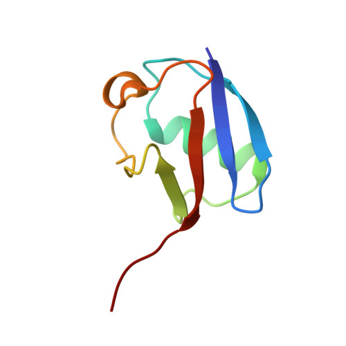
UbiquitinD [auth u]76Homo sapiensMutation(s): 0 
Gene Names: UBC
UniProt & NIH Common Fund Data Resources
Find proteins for P0CG48 (Homo sapiens)
Explore P0CG48 
Go to UniProtKB:  P0CG48
PHAROS:  P0CG48
GTEx:  ENSG00000150991 
Entity Groups
Sequence Clusters30% Identity50% Identity70% Identity90% Identity95% Identity100% Identity
UniProt GroupP0CG48
Sequence Annotations
Expand
Reference Sequence

Small Molecules

Modified Residues  1 Unique
IDChains TypeFormula2D DiagramParent
SEP
Query on SEP
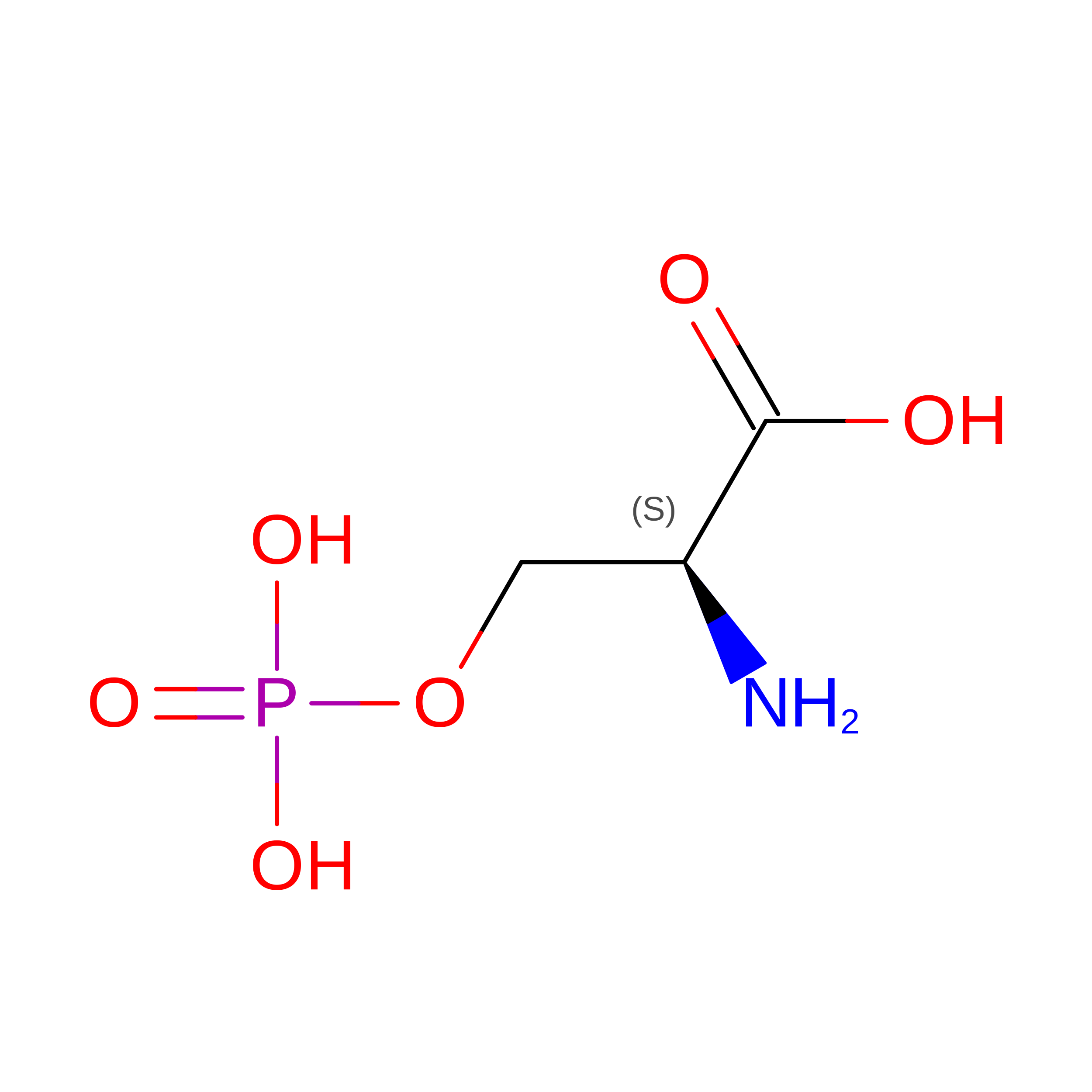
B [auth 2]L-PEPTIDE LINKINGC3 H8 N O6 PSER

Experimental Data & Validation

Experimental Data

  • Method: ELECTRON MICROSCOPY
  • Resolution: 3.40 Å
  • Aggregation State: PARTICLE 
  • Reconstruction Method: SINGLE PARTICLE 
EM Software:
TaskSoftware PackageVersion
MODEL REFINEMENTPHENIX

Structure Validation

View Full Validation Report


Entry History 

& Funding Information

Deposition Data


Funding OrganizationLocationGrant Number
Max Planck SocietyGermany--
European Research Council (ERC)European UnionH2020 789016-NEDD8Activate
German Research Foundation (DFG)GermanySCHU 3196/1-1

Revision History  (Full details and data files)

  • Version 1.0: 2024-01-03
    Type: Initial release
  • Version 1.1: 2024-01-24
    Changes: Derived calculations
  • Version 1.2: 2024-01-31
    Changes: Database references
  • Version 1.3: 2024-11-13
    Changes: Data collection, Structure summary
  • Version 1.4: 2025-07-09
    Changes: Data collection