8P9D

Crystal structure of p63-p73 heterotetramer (tetramerisation domain) in complex with darpin 1810 A2


Experimental Data Snapshot

  • Method: X-RAY DIFFRACTION
  • Resolution: 2.70 Å
  • R-Value Free: 0.252 
  • R-Value Work: 0.215 
  • R-Value Observed: 0.217 

wwPDB Validation   3D Report Full Report


This is version 1.1 of the entry. See complete history


Literature

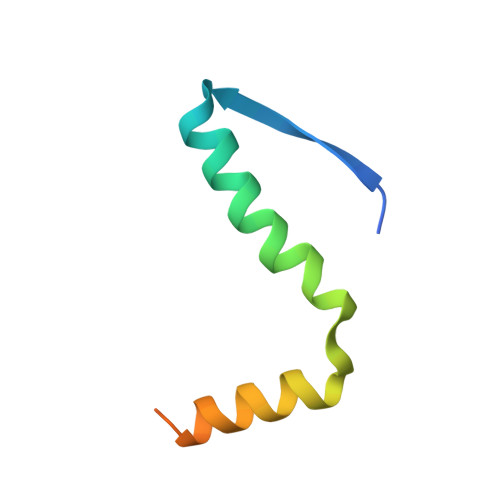
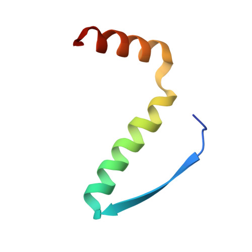
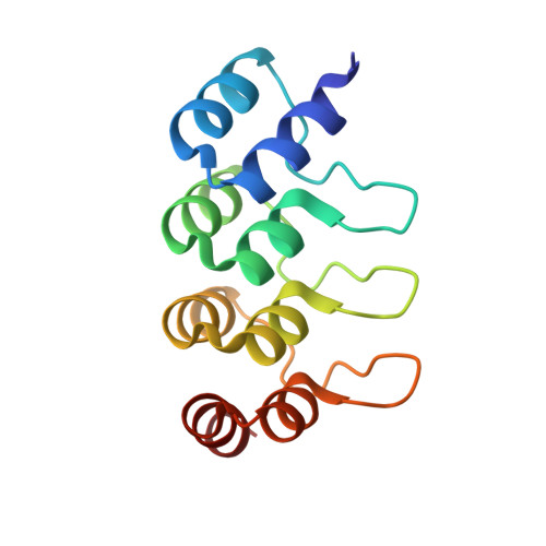
DARPins detect the formation of hetero-tetramers of p63 and p73 in epithelial tissues and in squamous cell carcinoma.

Strubel, A.Munick, P.Hartmann, O.Chaikuad, A.Dreier, B.Schaefer, J.V.Gebel, J.Osterburg, C.Tuppi, M.Schafer, B.Buck, V.Rosenfeldt, M.Knapp, S.Pluckthun, A.Diefenbacher, M.E.Dotsch, V.

(2023) Cell Death Dis 14: 674-674

  • DOI: https://doi.org/10.1038/s41419-023-06213-0
  • Primary Citation of Related Structures:  
    8P9C, 8P9D, 8P9E

  • PubMed Abstract: 

    The two p53 homologues p63 and p73 regulate transcriptional programs in epithelial tissues and several cell types in these tissues express both proteins. All members of the p53 family form tetramers in their active state through a dedicated oligomerization domain that structurally assembles as a dimer of dimers. The oligomerization domain of p63 and p73 share a high sequence identity, but the p53 oligomerization domain is more divergent and it lacks a functionally important C-terminal helix present in the other two family members. Based on these structural differences, p53 does not hetero-oligomerize with p63 or p73. In contrast, p63 and p73 form hetero-oligomers of all possible stoichiometries, with the hetero-tetramer built from a p63 dimer and a p73 dimer being thermodynamically more stable than the two homo-tetramers. This predicts that in cells expressing both proteins a p63 2 /p73 2 hetero-tetramer is formed. So far, the tools to investigate the biological function of this hetero-tetramer have been missing. Here we report the generation and characterization of Designed Ankyrin Repeat Proteins (DARPins) that bind with high affinity and selectivity to the p63 2 /p73 2 hetero-tetramer. Using these DARPins we were able to confirm experimentally the existence of this hetero-tetramer in epithelial mouse and human tissues and show that its level increases in squamous cell carcinoma.


  • Organizational Affiliation

    Institute of Biophysical Chemistry and Center for Biomolecular Magnetic Resonance, Goethe University, 60438, Frankfurt, Germany.


Macromolecules
Find similar proteins by:  (by identity cutoff)  |  3D Structure
Entity ID: 1
MoleculeChains Sequence LengthOrganismDetailsImage
Tumor protein 63
A, C
61Homo sapiensMutation(s): 0 
Gene Names: TP63KETP63P73HP73LTP73L
UniProt & NIH Common Fund Data Resources
Find proteins for Q9H3D4 (Homo sapiens)
Explore Q9H3D4 
Go to UniProtKB:  Q9H3D4
PHAROS:  Q9H3D4
GTEx:  ENSG00000073282 
Entity Groups  
Sequence Clusters30% Identity50% Identity70% Identity90% Identity95% Identity100% Identity
UniProt GroupQ9H3D4
Sequence Annotations
Expand
  • Reference Sequence
Find similar proteins by:  (by identity cutoff)  |  3D Structure
Entity ID: 2
MoleculeChains Sequence LengthOrganismDetailsImage
Tumor protein p73
B, D
50Homo sapiensMutation(s): 0 
Gene Names: TP73P73
UniProt & NIH Common Fund Data Resources
Find proteins for O15350 (Homo sapiens)
Explore O15350 
Go to UniProtKB:  O15350
PHAROS:  O15350
GTEx:  ENSG00000078900 
Entity Groups  
Sequence Clusters30% Identity50% Identity70% Identity90% Identity95% Identity100% Identity
UniProt GroupO15350
Sequence Annotations
Expand
  • Reference Sequence
Find similar proteins by:  (by identity cutoff)  |  3D Structure
Entity ID: 3
MoleculeChains Sequence LengthOrganismDetailsImage
Darpin 1810 A2
E, F
126Lama glamaMutation(s): 0 
Entity Groups  
Sequence Clusters30% Identity50% Identity70% Identity90% Identity95% Identity100% Identity
Sequence Annotations
Expand
  • Reference Sequence
Experimental Data & Validation

Experimental Data

  • Method: X-RAY DIFFRACTION
  • Resolution: 2.70 Å
  • R-Value Free: 0.252 
  • R-Value Work: 0.215 
  • R-Value Observed: 0.217 
  • Space Group: P 21 21 21
Unit Cell:
Length ( Å )Angle ( ˚ )
a = 40.729α = 90
b = 113.186β = 90
c = 135.227γ = 90
Software Package:
Software NamePurpose
REFMACrefinement
Aimlessdata scaling
XDSdata reduction
PHASERphasing

Structure Validation

View Full Validation Report


Entry History & Funding Information

Deposition Data


Funding OrganizationLocationGrant Number
Not funded--

Revision History  (Full details and data files)

  • Version 1.0: 2023-11-08
    Type: Initial release
  • Version 1.1: 2023-11-15
    Changes: Database references, Derived calculations, Source and taxonomy, Structure summary