8P99 | pdb_00008p99

SARS-CoV-2 S-protein:D614G mutant in 1-up conformation


Experimental Data Snapshot

  • Method: ELECTRON MICROSCOPY
  • Resolution: 3.40 Å
  • Aggregation State: PARTICLE 
  • Reconstruction Method: SINGLE PARTICLE 

wwPDB Validation 3D Report Full Report

Validation slider image for 8P99

This is version 1.1 of the entry. See complete history

Literature

C-2 Thiophenyl Tryptophan Trimers Inhibit Cellular Entry of SARS-CoV-2 through Interaction with the Viral Spike (S) Protein.

Gargantilla, M.Frances, C.Adhav, A.Forcada-Nadal, A.Martinez-Gualda, B.Marti-Mari, O.Lopez-Redondo, M.L.Melero, R.Marco-Marin, C.Gougeard, N.Espinosa, C.Rubio-Del-Campo, A.Ruiz-Partida, R.Hernandez-Sierra, M.D.P.Villamayor-Belinchon, L.Bravo, J.Llacer, J.L.Marina, A.Rubio, V.San-Felix, A.Geller, R.Perez-Perez, M.J.

(2023) J Med Chem 66: 10432-10457

  • DOI: https://doi.org/10.1021/acs.jmedchem.3c00576
  • Primary Citation Related Structures: 
    8P99, 8P9Y

  • PubMed Abstract: 

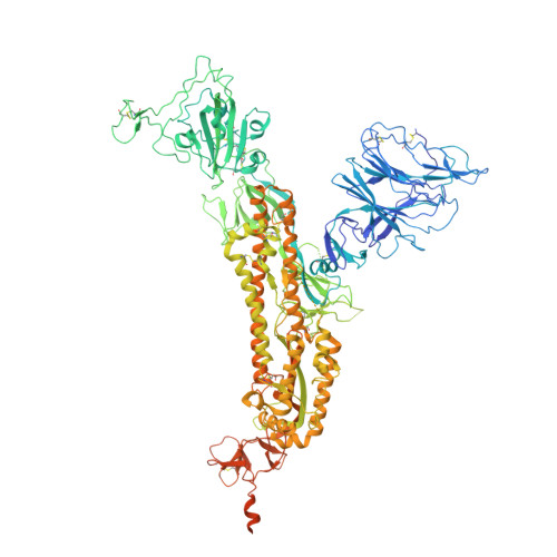
    Severe acute respiratory syndrome coronavirus 2 (SARS-CoV-2) causes COVID-19, by infecting cells via the interaction of its spike protein (S) with the primary cell receptor angiotensin-converting enzyme (ACE2). To search for inhibitors of this key step in viral infection, we screened an in-house library of multivalent tryptophan derivatives. Using VSV-S pseudoparticles, we identified compound 2 as a potent entry inhibitor lacking cellular toxicity. Chemical optimization of 2 rendered compounds 63 and 65 , which also potently inhibited genuine SARS-CoV-2 cell entry. Thermofluor and microscale thermophoresis studies revealed their binding to S and to its isolated receptor binding domain (RBD), interfering with the interaction with ACE2. High-resolution cryoelectron microscopy structure of S, free or bound to 2 , shed light on cell entry inhibition mechanisms by these compounds. Overall, this work identifies and characterizes a new class of SARS-CoV-2 entry inhibitors with clear potential for preventing and/or fighting COVID-19.


  • Organizational Affiliation
    • Instituto de Química Médica (IQM, CSIC), c/Juan de la Cierva 3, Madrid 28006, Spain.

Macromolecule Content 

  • Total Structure Weight: 432.27 kDa 
  • Atom Count: 25,713 
  • Modeled Residue Count: 3,219 
  • Deposited Residue Count: 3,810 
  • Unique protein chains: 1

Macromolecules

Find similar proteins by:|  3D Structure
Entity ID: 1
MoleculeChains  Sequence LengthOrganismDetailsImage
Spike protein S1,Spike glycoprotein
A, B, C
1,270Severe acute respiratory syndrome coronavirus 2Mutation(s): 4 
Gene Names: S2
UniProt
Find proteins for P0DTC2 (Severe acute respiratory syndrome coronavirus 2)
Explore P0DTC2 
Go to UniProtKB:  P0DTC2
Entity Groups
Sequence Clusters30% Identity50% Identity70% Identity90% Identity95% Identity100% Identity
UniProt GroupP0DTC2
Glycosylation
Glycosylation Sites: 8Go to GlyGen: P0DTC2-1
Sequence Annotations
Expand
Reference Sequence

Oligosaccharides

Help  
Entity ID: 2
MoleculeChains Length2D Diagram GlycosylationD Interactions
2-acetamido-2-deoxy-beta-D-glucopyranose-(1-4)-2-acetamido-2-deoxy-beta-D-glucopyranose
D [auth E],
E [auth F],
F [auth G],
G [auth I],
H [auth J],
D [auth E],
E [auth F],
F [auth G],
G [auth I],
H [auth J],
I [auth K],
J [auth L],
K [auth N],
L [auth O],
M [auth P]
2N-Glycosylation
Glycosylation Resources
GlyTouCan: G42666HT
GlyCosmos: G42666HT
GlyGen: G42666HT

Small Molecules

Ligands 1 Unique
IDChains Name / Formula / InChI Key2D Diagram3D Interactions
NAG

Query on NAG


Download:Ideal Coordinates CCD File
AA [auth B]
BA [auth B]
CA [auth B]
DA [auth B]
EA [auth C]
AA [auth B],
BA [auth B],
CA [auth B],
DA [auth B],
EA [auth C],
FA [auth C],
GA [auth C],
HA [auth C],
IA [auth C],
JA [auth C],
KA [auth C],
N [auth A],
O [auth A],
P [auth A],
Q [auth A],
R [auth A],
S [auth A],
T [auth A],
U [auth A],
V [auth B],
W [auth B],
X [auth B],
Y [auth B],
Z [auth B]
2-acetamido-2-deoxy-beta-D-glucopyranose
C8 H15 N O6
OVRNDRQMDRJTHS-FMDGEEDCSA-N

Experimental Data & Validation

Experimental Data

  • Method: ELECTRON MICROSCOPY
  • Resolution: 3.40 Å
  • Aggregation State: PARTICLE 
  • Reconstruction Method: SINGLE PARTICLE 
EM Software:
TaskSoftware PackageVersion
MODEL REFINEMENTREFMAC5.8.0158

Structure Validation

View Full Validation Report


Entry History 

& Funding Information

Deposition Data


Funding OrganizationLocationGrant Number
European CommissionEuropean UnionEU 2020/2094
Spanish Ministry of Science, Innovation, and UniversitiesSpainPID2020 116880GB-I00
Spanish Ministry of Science, Innovation, and UniversitiesSpainPID2020 120322RB-C21
Spanish National Research CouncilSpain202080E110

Revision History  (Full details and data files)

  • Version 1.0: 2023-09-27
    Type: Initial release
  • Version 1.1: 2024-11-13
    Changes: Data collection, Structure summary