8P6Q

Racemic structure of TNFR1 cysteine-rich domain

  • Classification: CYTOKINE
  • Organism(s): Homo sapiens
  • Mutation(s): No 

  • Deposited: 2023-05-28 Released: 2024-01-24 
  • Deposition Author(s): Lander, A.J., Jin, Y., Luk, L.Y.P.
  • Funding Organization(s): Engineering and Physical Sciences Research Council, Wellcome Trust, Biotechnology and Biological Sciences Research Council (BBSRC)

Experimental Data Snapshot

  • Method: X-RAY DIFFRACTION
  • Resolution: 1.40 Å
  • R-Value Free: 0.251 
  • R-Value Work: 0.200 
  • R-Value Observed: 0.203 

wwPDB Validation   3D Report Full Report


This is version 1.1 of the entry. See complete history


Literature

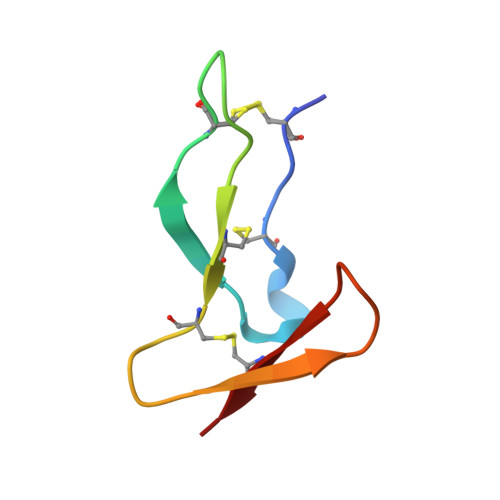
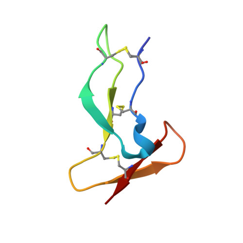
Deciphering the Synthetic and Refolding Strategy of a Cysteine-Rich Domain in the Tumor Necrosis Factor Receptor (TNF-R) for Racemic Crystallography Analysis and d-Peptide Ligand Discovery.

Lander, A.J.Kong, Y.Jin, Y.Wu, C.Luk, L.Y.P.

(2024) ACS Bio Med Chem Au 4: 68-76

  • DOI: https://doi.org/10.1021/acsbiomedchemau.3c00060
  • Primary Citation of Related Structures:  
    8P6Q

  • PubMed Abstract: 

    Many cell-surface receptors are promising targets for chemical synthesis because of their critical roles in disease development. This synthetic approach enables investigations by racemic protein crystallography and ligand discovery by mirror-image methodologies. However, due to their complex nature, the chemical synthesis of a receptor can be a significant challenge. Here, we describe the chemical synthesis and folding of a central, cysteine-rich domain of the cell-surface receptor tumor necrosis factor 1 which is integral to binding of the cytokine TNF-α, namely, TNFR-1 CRD2. Racemic protein crystallography at 1.4 Å confirmed that the native binding conformation was preserved, and TNFR-1 CRD2 maintained its capacity to bind to TNF-α ( K D ≈ 7 nM). Encouraged by this discovery, we carried out mirror-image phage display using the enantiomeric receptor mimic and identified a d-peptide ligand for TNFR-1 CRD2 ( K D = 1 μM). This work demonstrated that cysteine-rich domains, including the central domains, can be chemically synthesized and used as mimics for investigations.


  • Organizational Affiliation

    School of Chemistry, Cardiff University, Main Building, Park Place, Cardiff CF10 3AT, U.K.


Macromolecules
Find similar proteins by:  (by identity cutoff)  |  3D Structure
Entity ID: 1
MoleculeChains Sequence LengthOrganismDetailsImage
Tumor necrosis factor-binding protein 145Homo sapiensMutation(s): 0 
UniProt & NIH Common Fund Data Resources
Find proteins for P19438 (Homo sapiens)
Explore P19438 
Go to UniProtKB:  P19438
PHAROS:  P19438
GTEx:  ENSG00000067182 
Entity Groups  
Sequence Clusters30% Identity50% Identity70% Identity90% Identity95% Identity100% Identity
UniProt GroupP19438
Sequence Annotations
Expand
  • Reference Sequence
Find similar proteins by:  (by identity cutoff)  |  3D Structure
Entity ID: 2
MoleculeChains Sequence LengthOrganismDetailsImage
D-TNFR-1 CRD245Homo sapiensMutation(s): 0 
Sequence Annotations
Expand
  • Reference Sequence
Experimental Data & Validation

Experimental Data

  • Method: X-RAY DIFFRACTION
  • Resolution: 1.40 Å
  • R-Value Free: 0.251 
  • R-Value Work: 0.200 
  • R-Value Observed: 0.203 
  • Space Group: P 1 21 1
Unit Cell:
Length ( Å )Angle ( ˚ )
a = 20.42α = 90
b = 50.49β = 92.94
c = 46.21γ = 90
Software Package:
Software NamePurpose
REFMACrefinement
xia2data reduction
Aimlessdata scaling
MOLREPphasing
Cootmodel building

Structure Validation

View Full Validation Report


Entry History & Funding Information

Deposition Data


Funding OrganizationLocationGrant Number
Engineering and Physical Sciences Research CouncilUnited Kingdom2107414
Wellcome TrustUnited Kingdom218568/Z/19/Z
Biotechnology and Biological Sciences Research Council (BBSRC)United KingdomBB/T015799/1

Revision History  (Full details and data files)

  • Version 1.0: 2024-01-24
    Type: Initial release
  • Version 1.1: 2024-03-06
    Changes: Database references