8P5M | pdb_00008p5m

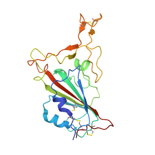
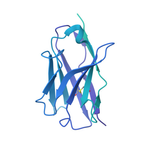
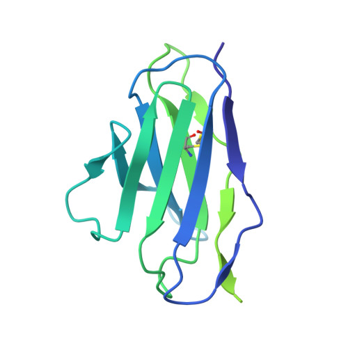
SARS-CoV-2 Spike RBD in complex with Mab-23 (Fab)


Experimental Data Snapshot

  • Method: ELECTRON MICROSCOPY
  • Resolution: 2.80 Å
  • Aggregation State: PARTICLE 
  • Reconstruction Method: SINGLE PARTICLE 

Starting Model: experimental
View more details

wwPDB Validation 3D Report Full Report

Validation slider image for 8P5M

This is version 1.3 of the entry. See complete history. 

Literature

Multi-compartmental diversification of neutralizing antibody lineages dissected in SARS-CoV-2 spike-immunized macaques.

Mandolesi, M., Das, H., de Vries, L., Yang, Y., Kim, C., Dhinakaran, M., Castro Dopico, X., Fischbach, J., Kim, S., Guryleva, M.V., Adori, M., Chernyshev, M., Stalmarck, A., Hanke, L., McInerney, G.M., Sheward, D.J., Corcoran, M., Hallberg, B.M., Murrell, B., Karlsson Hedestam, G.B.

(2024) Nat Commun 15: 6338-6338

  • DOI: https://doi.org/10.1038/s41467-024-50286-0
  • Primary Citation Related Structures: 
    8P5M, 8Q5Y

  • PubMed Abstract: 

    The continued evolution of SARS-CoV-2 underscores the need to understand qualitative aspects of the humoral immune response elicited by spike immunization. Here, we combine monoclonal antibody (mAb) isolation with deep B cell receptor (BCR) repertoire sequencing of rhesus macaques immunized with prefusion-stabilized spike glycoprotein. Longitudinal tracing of spike-sorted B cell lineages in multiple immune compartments demonstrates increasing somatic hypermutation and broad dissemination of vaccine-elicited B cells in draining and non-draining lymphoid compartments, including the bone marrow, spleen and, most notably, periaortic lymph nodes. Phylogenetic analysis of spike-specific monoclonal antibody lineages identified through deep repertoire sequencing delineates extensive intra-clonal diversification that shaped neutralizing activity. Structural analysis of the spike in complex with a broadly neutralizing mAb provides a molecular basis for the observed differences in neutralization breadth between clonally related antibodies. Our findings highlight that immunization leads to extensive intra-clonal B cell evolution where members of the same lineage can both retain the original epitope specificity and evolve to recognize additional spike variants not previously encountered.


  • Organizational Affiliation: 
    • Department of Microbiology, Tumor and Cell Biology, Karolinska Institutet, Stockholm, Sweden. marco.mandolesi.job@gmail.com.

Macromolecule Content 

  • Total Structure Weight: 93.74 kDa 
  • Atom Count: 3,258 
  • Modeled Residue Count: 419 
  • Deposited Residue Count: 852 
  • Unique protein chains: 3

Macromolecules

Find similar proteins by:|  3D Structure
Entity ID: 1
MoleculeChains  Sequence LengthOrganismDetailsImage
Spike protein S1A [auth I]196Severe acute respiratory syndrome coronavirus 2Mutation(s): 0 
Gene Names: S, 2
UniProt
Find proteins for P0DTC2 (Severe acute respiratory syndrome coronavirus 2)
Explore P0DTC2 
Go to UniProtKB:  P0DTC2
Entity Groups
Sequence Clusters30% Identity50% Identity70% Identity90% Identity95% Identity100% Identity
UniProt GroupP0DTC2
Sequence Annotations
Expand
Reference Sequence
Find similar proteins by:|  3D Structure
Entity ID: 2
MoleculeChains  Sequence LengthOrganismDetailsImage
Mab-23 (Heavy chain variable domain)B [auth G]442Homo sapiensMutation(s): 0 
Entity Groups
Sequence Clusters30% Identity50% Identity70% Identity90% Identity95% Identity100% Identity
Sequence Annotations
Expand
Reference Sequence
Find similar proteins by:|  3D Structure
Entity ID: 3
MoleculeChains  Sequence LengthOrganismDetailsImage
Mab-23 (Light chain variable domain)C [auth L]214Homo sapiensMutation(s): 0 
Entity Groups
Sequence Clusters30% Identity50% Identity70% Identity90% Identity95% Identity100% Identity
Sequence Annotations
Expand
Reference Sequence

Experimental Data & Validation

Experimental Data

  • Method: ELECTRON MICROSCOPY
  • Resolution: 2.80 Å
  • Aggregation State: PARTICLE 
  • Reconstruction Method: SINGLE PARTICLE 
EM Software:
TaskSoftware PackageVersion
MODEL REFINEMENTPHENIX1.19
RECONSTRUCTIONcryoSPARC3.2.0
RECONSTRUCTIONcryoSPARC4.1.1

Structure Validation

View Full Validation Report


Entry History 

& Funding Information

Deposition Data


Funding OrganizationLocationGrant Number
Not funded--

Revision History  (Full details and data files)

  • Version 1.0: 2024-06-05
    Type: Initial release
  • Version 1.1: 2024-09-25
    Changes: Data collection, Source and taxonomy
  • Version 1.2: 2024-10-23
    Changes: Data collection, Structure summary
  • Version 1.3: 2024-12-18
    Changes: Data collection, Database references