Structural and functional studies of Arabidopsis thaliana glutamate dehydrogenase isoform 2 demonstrate enzyme dynamics and identify its calcium binding site.
Grzechowiak, M., Sliwiak, J., Jaskolski, M., Ruszkowski, M.(2023) Plant Physiol Biochem 201: 107895-107895
- PubMed: 37478728 Search on PubMed
- DOI: https://doi.org/10.1016/j.plaphy.2023.107895
- Primary Citation Related Structures:
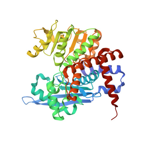
8OWM, 8OWN - PubMed Abstract:
Glutamate dehydrogenase (GDH) is an enzyme at the crossroad of plant nitrogen and carbon metabolism. GDH catalyzes the conversion of 2-oxoglutarate into glutamate (2OG → Glu), utilizing ammonia as cosubstrate and NADH as coenzyme. The GDH reaction is reversible, meaning that the NAD + -dependent reaction (Glu → 2OG) releases ammonia. In Arabidopsis thaliana, three GDH isoforms exist, AtGDH1, AtGDH2, and AtGDH3. The subject of this work is AtGDH2. Previous reports have suggested that enzymes homologous to AtGDH2 contain a calcium-binding EF-hand motif located in the coenzyme binding domain. Here, we show that while AtGDH2 indeed does bind calcium, the binding occurs elsewhere and the region predicted to be the EF-hand motif has a completely different structure. As the true calcium binding site is > 20 Å away from the active site, it seems to play a structural, rather than catalytic role. We also performed comparative kinetic characterization of AtGDH1 and AtGDH2 using spectroscopic methods and isothermal titration calorimetry, to note that the isoenzymes generally exhibit similar behavior, with calcium having only a minor effect. However, the spatial and temporal changes in the gene expression profiles of the three AtGDH genes point to AtGDH2 as the most prevalent isoform.
- Department of Structural Biology of Eukaryotes, Institute of Bioorganic Chemistry, Polish Academy of Sciences, Poznan, 61-704, Poland.
Organizational Affiliation: