Download Mendeley
Multimeric structure of a subfamily III haloalkane dehalogenase-like enzyme solved by combination of cryo-EM and x-ray crystallography.
Chmelova, K., Gao, T., Polak, M., Schenkmayerova, A., Croll, T.I., Shaikh, T.R., Skarupova, J., Chaloupkova, R., Diederichs, K., Read, R.J., Damborsky, J., Novacek, J., Marek, M.(2023) Protein Sci 32: e4751-e4751
- PubMed: 37574754 Search on PubMedSearch on PubMed Central
- DOI: https://doi.org/10.1002/pro.4751
- Primary Citation Related Structures:
8CKP, 8OOH - PubMed Abstract:
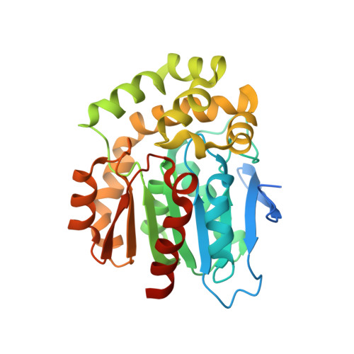
Haloalkane dehalogenase (HLD) enzymes employ an S N 2 nucleophilic substitution mechanism to erase halogen substituents in diverse organohalogen compounds. Subfamily I and II HLDs are well-characterized enzymes, but the mode and purpose of multimerization of subfamily III HLDs are unknown. Here we probe the structural organization of DhmeA, a subfamily III HLD-like enzyme from the archaeon Haloferax mediterranei, by combining cryo-electron microscopy (cryo-EM) and x-ray crystallography. We show that full-length wild-type DhmeA forms diverse quaternary structures, ranging from small oligomers to large supramolecular ring-like assemblies of various sizes and symmetries. We optimized sample preparation steps, enabling three-dimensional reconstructions of an oligomeric species by single-particle cryo-EM. Moreover, we engineered a crystallizable mutant (DhmeA ΔGG ) that provided diffraction-quality crystals. The 3.3 Å crystal structure reveals that DhmeA ΔGG forms a ring-like 20-mer structure with outer and inner diameter of ~200 and ~80 Å, respectively. An enzyme homodimer represents a basic repeating building unit of the crystallographic ring. Three assembly interfaces (dimerization, tetramerization, and multimerization) were identified to form the supramolecular ring that displays a negatively charged exterior, while its interior part harboring catalytic sites is positively charged. Localization and exposure of catalytic machineries suggest a possible processing of large negatively charged macromolecular substrates.
- Loschmidt Laboratories, Department of Experimental Biology and RECETOX, Faculty of Science, Masaryk University, Brno, Czech Republic.
Organizational Affiliation: