Structural and functional analysis of aquaporin-2 mutants involved in nephrogenic diabetes insipidus.
Hagstromer, C.J., Hyld Steffen, J., Kreida, S., Al-Jubair, T., Frick, A., Gourdon, P., Tornroth-Horsefield, S.(2023) Sci Rep 13: 14674-14674
- PubMed: 37674034 Search on PubMedSearch on PubMed Central
- DOI: https://doi.org/10.1038/s41598-023-41616-1
- Primary Citation Related Structures:
8GHJ, 8OEE - PubMed Abstract:
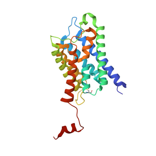
Aquaporins are water channels found in the cell membrane, where they allow the passage of water molecules in and out of the cells. In the kidney collecting duct, arginine vasopressin-dependent trafficking of aquaporin-2 (AQP2) fine-tunes reabsorption of water from pre-urine, allowing precise regulation of the final urine volume. Point mutations in the gene for AQP2 may disturb this process and lead to nephrogenic diabetes insipidus (NDI), whereby patients void large volumes of highly hypo-osmotic urine. In recessive NDI, mutants of AQP2 are retained in the endoplasmic reticulum due to misfolding. Here we describe the structural and functional characterization of three AQP2 mutations associated with recessive NDI: T125M and T126M, situated close to a glycosylation site and A147T in the transmembrane region. Using a proteoliposome assay, we show that all three mutants permit the transport of water. The crystal structures of T125M and T126M together with biophysical characterization of all three mutants support that they retain the native structure, but that there is a significant destabilization of A147T. Our work provides unique molecular insights into the mechanisms behind recessive NDI as well as deepens our understanding of how misfolded proteins are recognized by the ER quality control system.
- Department of Biochemistry and Structural Biology, Lund University, Lund, Sweden.
Organizational Affiliation: