8KDX

Tau-S214 Phosphorylation Inhibits Fyn Kinase Interaction and Increases the Decay Time of NMDAR-mediated Current


Experimental Data Snapshot

  • Method: X-RAY DIFFRACTION
  • Resolution: 1.01 Å
  • R-Value Free: 0.185 
  • R-Value Work: 0.173 

wwPDB Validation   3D Report Full Report


This is version 1.0 of the entry. See complete history


Literature

Tau-S214 Phosphorylation Inhibits Fyn Kinase Interaction and Increases the Decay Time of NMDAR-mediated Current.

Jos, S.Poulose, R.Kambaru, A.Gogoi, H.Dalavaikodihalli Nanjaiah, N.Padmanabhan, B.Mehta, B.Padavattan, S.

(2024) J Mol Biol 436: 168445-168445

  • DOI: https://doi.org/10.1016/j.jmb.2024.168445
  • Primary Citation of Related Structures:  
    8KDX

  • PubMed Abstract: 

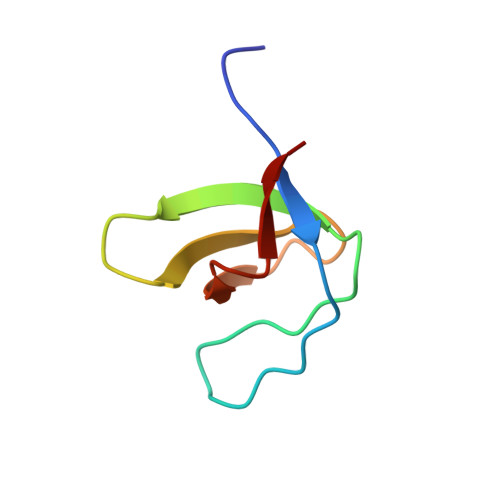
    Fyn kinase SH3 domain interaction with PXXP motif in the Tau protein is implicated in AD pathology and is central to NMDAR function. Among seven PXXP motifs localized in proline-rich domain of Tau protein, tandem 5th and 6th PXXP motifs are critical to Fyn-SH3 domain interaction. Here, we report the crystal structure of Fyn-SH3 -Tau (207-221) peptide consisting of 5th and 6th PXXP motif complex to 1.01 Å resolution. Among five AD-specific phosphorylation sites encompassing the 5th and 6th PXXP motifs, only S214 residue showed interaction with SH3 domain. Biophysical studies showed that Tau (207-221) with S214-phosphorylation (pS214) inhibits its interaction with Fyn-SH3 domain. The individual administration of Tau (207-221) with/without pS214 peptides to a single neuron increased the decay time of evoked NMDA current response. Recordings of spontaneous NMDA EPSCs at +40 mV indicate an increase in frequency and amplitude of events for the Tau (207-221) peptide. Conversely, the Tau (207-221) with pS214 peptide exhibited a noteworthy amplitude increase alongside a prolonged decay time. These outcomes underscore the distinctive modalities of action associated with each peptide in the study. Overall, this study provides insights into how Tau (207-221) with/without pS214 affects the molecular framework of NMDAR signaling, indicating its involvement in Tau-related pathogenesis.


  • Organizational Affiliation

    Department of Biophysics, National Institute of Mental Health and Neurosciences, Bangalore 560029, India.


Macromolecules
Find similar proteins by:  (by identity cutoff)  |  3D Structure
Entity ID: 1
MoleculeChains Sequence LengthOrganismDetailsImage
Tyrosine-protein kinase Fyn61Homo sapiensMutation(s): 0 
Gene Names: FYN
EC: 2.7.10.2
UniProt & NIH Common Fund Data Resources
Find proteins for P06241 (Homo sapiens)
Explore P06241 
Go to UniProtKB:  P06241
PHAROS:  P06241
GTEx:  ENSG00000010810 
Entity Groups  
Sequence Clusters30% Identity50% Identity70% Identity90% Identity95% Identity100% Identity
UniProt GroupP06241
Sequence Annotations
Expand
  • Reference Sequence

Find similar proteins by:  Sequence   |   3D Structure  

Entity ID: 2
MoleculeChains Sequence LengthOrganismDetailsImage
Microtubule-associated protein tau15Homo sapiensMutation(s): 0 
UniProt & NIH Common Fund Data Resources
Find proteins for P10636 (Homo sapiens)
Explore P10636 
Go to UniProtKB:  P10636
PHAROS:  P10636
GTEx:  ENSG00000186868 
Entity Groups  
Sequence Clusters30% Identity50% Identity70% Identity90% Identity95% Identity100% Identity
UniProt GroupP10636
Sequence Annotations
Expand
  • Reference Sequence
Experimental Data & Validation

Experimental Data

  • Method: X-RAY DIFFRACTION
  • Resolution: 1.01 Å
  • R-Value Free: 0.185 
  • R-Value Work: 0.173 
  • Space Group: P 21 21 21
Unit Cell:
Length ( Å )Angle ( ˚ )
a = 31.152α = 90
b = 35.558β = 90
c = 73.169γ = 90
Software Package:
Software NamePurpose
REFMACrefinement
iMOSFLMdata reduction
Aimlessdata scaling
PHASERphasing

Structure Validation

View Full Validation Report


Entry History & Funding Information

Deposition Data


Funding OrganizationLocationGrant Number
Not fundedIndia--

Revision History  (Full details and data files)

  • Version 1.0: 2024-02-28
    Type: Initial release