8KDN | pdb_00008kdn

Structure of LAT1-CD98hc in complex with L-Phe, focused on TMD


Experimental Data Snapshot

  • Method: ELECTRON MICROSCOPY
  • Resolution: 4.12 Å
  • Aggregation State: PARTICLE 
  • Reconstruction Method: SINGLE PARTICLE 

wwPDB Validation 3D Report Full Report

Validation slider image for 8KDN

This is version 1.1 of the entry. See complete history

Literature

Structural basis of anticancer drug recognition and amino acid transport by LAT1.

Lee, Y.Jin, C.Ohgaki, R.Xu, M.Ogasawara, S.Warshamanage, R.Yamashita, K.Murshudov, G.Nureki, O.Murata, T.Kanai, Y.

(2025) Nat Commun 16: 1635-1635

  • DOI: https://doi.org/10.1038/s41467-025-56903-w
  • Primary Citation Related Structures: 
    8KDD, 8KDF, 8KDG, 8KDH, 8KDI, 8KDJ, 8KDN, 8KDO, 8KDP

  • PubMed Abstract: 

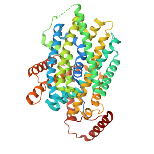
    LAT1 (SLC7A5) transports large neutral amino acids and plays pivotal roles in cancer proliferation, immune response and drug delivery. Despite recent advances in structural understanding of LAT1, how it discriminates substrates and inhibitors including the clinically relevant drugs remains elusive. Here we report six structures of LAT1 across three conformations with bound ligands, elucidating its substrate transport and inhibitory mechanisms. JPH203 (also known as nanvuranlat or KYT-0353), an anticancer drug in clinical trials, traps LAT1 in an outward-facing state with a U-shaped conformer, with its amino-phenylbenzoxazol moiety pushing against transmembrane helix 3 (TM3) and bending TM10. Physiological substrates like ʟ-Phe lack such effects, whereas melphalan poses steric hindrance, explaining its inhibitory activity. The "classical" system L inhibitor BCH induces an occluded state critical for transport, confirming its substrate-like behavior. These findings provide a structural basis for substrate recognition and inhibition of LAT1, guiding future drug design.


  • Organizational Affiliation
    • Department of Structural Biology, Max Planck Institute of Biophysics, 60438, Frankfurt, Germany. yongchan.lee@biophys.mpg.de.

Macromolecule Content 

  • Total Structure Weight: 124.28 kDa 
  • Atom Count: 3,930 
  • Modeled Residue Count: 506 
  • Deposited Residue Count: 1,146 
  • Unique protein chains: 2

Macromolecules

Find similar proteins by:|  3D Structure
Entity ID: 1
MoleculeChains  Sequence LengthOrganismDetailsImage
4F2 cell-surface antigen heavy chain631Homo sapiensMutation(s): 0 
Gene Names: SLC3A2MDU1
Membrane Entity: Yes 
UniProt & NIH Common Fund Data Resources
Find proteins for P08195 (Homo sapiens)
Explore P08195 
Go to UniProtKB:  P08195
PHAROS:  P08195
GTEx:  ENSG00000168003 
Entity Groups
Sequence Clusters30% Identity50% Identity70% Identity90% Identity95% Identity100% Identity
UniProt GroupP08195
Sequence Annotations
Expand
Reference Sequence
Find similar proteins by:|  3D Structure
Entity ID: 2
MoleculeChains  Sequence LengthOrganismDetailsImage
Large neutral amino acids transporter small subunit 1515Homo sapiensMutation(s): 0 
Gene Names: SLC7A5CD98LCLAT1MPE16
Membrane Entity: Yes 
UniProt & NIH Common Fund Data Resources
Find proteins for Q01650 (Homo sapiens)
Explore Q01650 
Go to UniProtKB:  Q01650
PHAROS:  Q01650
GTEx:  ENSG00000103257 
Entity Groups
Sequence Clusters30% Identity50% Identity70% Identity90% Identity95% Identity100% Identity
UniProt GroupQ01650
Sequence Annotations
Expand
Reference Sequence

Small Molecules

Ligands 1 Unique
IDChains Name / Formula / InChI Key2D Diagram3D Interactions
PHE
(Subject of Investigation/LOI)

Query on PHE


Download:Ideal Coordinates CCD File
C [auth B]PHENYLALANINE
C9 H11 N O2
COLNVLDHVKWLRT-QMMMGPOBSA-N

Experimental Data & Validation

Experimental Data

  • Method: ELECTRON MICROSCOPY
  • Resolution: 4.12 Å
  • Aggregation State: PARTICLE 
  • Reconstruction Method: SINGLE PARTICLE 

Structure Validation

View Full Validation Report


Entry History 

& Funding Information

Deposition Data

  • Released Date: 2025-02-12 
  • Deposition Author(s): Lee, Y.

Funding OrganizationLocationGrant Number
Max Planck SocietyGermany--

Revision History  (Full details and data files)

  • Version 1.0: 2025-02-12
    Type: Initial release
  • Version 1.1: 2025-02-26
    Changes: Data collection, Database references