8K6Y

Serial femtosecond crystallography structure of photo dissociated CO from ba3- type cytochrome c oxidase determined by extrapolation method


Experimental Data Snapshot

  • Method: X-RAY DIFFRACTION
  • Resolution: 2.00 Å
  • R-Value Free: 0.435 
  • R-Value Work: 0.396 

wwPDB Validation   3D Report Full Report


Ligand Structure Quality Assessment 


This is version 1.1 of the entry. See complete history


Literature

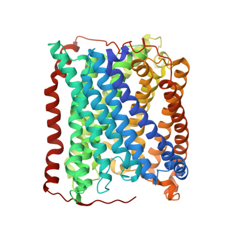
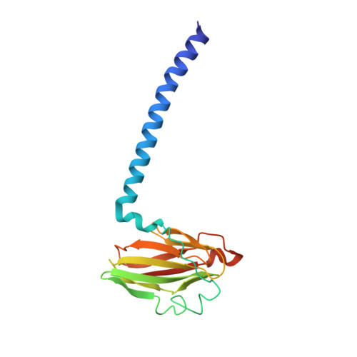
Time-resolved serial crystallography to track the dynamics of carbon monoxide in the active site of cytochrome c oxidase.

Safari, C.Ghosh, S.Andersson, R.Johannesson, J.Bath, P.Uwangue, O.Dahl, P.Zoric, D.Sandelin, E.Vallejos, A.Nango, E.Tanaka, R.Bosman, R.Borjesson, P.Dunevall, E.Hammarin, G.Ortolani, G.Panman, M.Tanaka, T.Yamashita, A.Arima, T.Sugahara, M.Suzuki, M.Masuda, T.Takeda, H.Yamagiwa, R.Oda, K.Fukuda, M.Tosha, T.Naitow, H.Owada, S.Tono, K.Nureki, O.Iwata, S.Neutze, R.Branden, G.

(2023) Sci Adv 9: eadh4179-eadh4179

  • DOI: https://doi.org/10.1126/sciadv.adh4179
  • Primary Citation of Related Structures:  
    8AJZ, 8K65, 8K6Y

  • PubMed Abstract: 

    Cytochrome c oxidase (C c O) is part of the respiratory chain and contributes to the electrochemical membrane gradient in mitochondria as well as in many bacteria, as it uses the energy released in the reduction of oxygen to pump protons across an energy-transducing biological membrane. Here, we use time-resolved serial femtosecond crystallography to study the structural response of the active site upon flash photolysis of carbon monoxide (CO) from the reduced heme a 3 of ba 3 -type C c O. In contrast with the aa 3 -type enzyme, our data show how CO is stabilized on Cu B through interactions with a transiently ordered water molecule. These results offer a structural explanation for the extended lifetime of the Cu B -CO complex in ba 3 -type C c O and, by extension, the extremely high oxygen affinity of the enzyme.


  • Organizational Affiliation

    Department of Chemistry and Molecular Biology, University of Gothenburg, Box 462, SE-40530 Gothenburg, Sweden.


Macromolecules
Find similar proteins by:  (by identity cutoff)  |  3D Structure
Entity ID: 1
MoleculeChains Sequence LengthOrganismDetailsImage
Cytochrome c oxidase subunit 1569Thermus thermophilus HB8Mutation(s): 0 
Gene Names: cbaATTHA1135
EC: 7.1.1.9
Membrane Entity: Yes 
UniProt
Find proteins for Q5SJ79 (Thermus thermophilus (strain ATCC 27634 / DSM 579 / HB8))
Explore Q5SJ79 
Go to UniProtKB:  Q5SJ79
Entity Groups  
Sequence Clusters30% Identity50% Identity70% Identity90% Identity95% Identity100% Identity
UniProt GroupQ5SJ79
Sequence Annotations
Expand
  • Reference Sequence
Find similar proteins by:  (by identity cutoff)  |  3D Structure
Entity ID: 2
MoleculeChains Sequence LengthOrganismDetailsImage
Cytochrome c oxidase subunit 2168Thermus thermophilus HB8Mutation(s): 0 
Gene Names: cbaBctaCTTHA1134
EC: 7.1.1.9
Membrane Entity: Yes 
UniProt
Find proteins for Q5SJ80 (Thermus thermophilus (strain ATCC 27634 / DSM 579 / HB8))
Explore Q5SJ80 
Go to UniProtKB:  Q5SJ80
Entity Groups  
Sequence Clusters30% Identity50% Identity70% Identity90% Identity95% Identity100% Identity
UniProt GroupQ5SJ80
Sequence Annotations
Expand
  • Reference Sequence
Find similar proteins by:  (by identity cutoff)  |  3D Structure
Entity ID: 3
MoleculeChains Sequence LengthOrganismDetailsImage
Cytochrome c oxidase polypeptide 2A34Thermus thermophilus HB8Mutation(s): 0 
Gene Names: cbaDTTHA1133
EC: 7.1.1.9
Membrane Entity: Yes 
UniProt
Find proteins for P82543 (Thermus thermophilus (strain ATCC 27634 / DSM 579 / HB8))
Explore P82543 
Go to UniProtKB:  P82543
Entity Groups  
Sequence Clusters30% Identity50% Identity70% Identity90% Identity95% Identity100% Identity
UniProt GroupP82543
Sequence Annotations
Expand
  • Reference Sequence
Small Molecules
Ligands 6 Unique
IDChains Name / Formula / InChI Key2D Diagram3D Interactions
HAS (Subject of Investigation/LOI)
Query on HAS

Download Ideal Coordinates CCD File 
F [auth A]HEME-AS
C54 H64 Fe N4 O6
PDYODZVCODUKFH-ZOMLSHGTSA-L
HEM
Query on HEM

Download Ideal Coordinates CCD File 
E [auth A]PROTOPORPHYRIN IX CONTAINING FE
C34 H32 Fe N4 O4
KABFMIBPWCXCRK-RGGAHWMASA-L
OLC
Query on OLC

Download Ideal Coordinates CCD File 
G [auth A]
H [auth A]
I [auth A]
J [auth A]
K [auth A]
G [auth A],
H [auth A],
I [auth A],
J [auth A],
K [auth A],
L [auth A],
M [auth A],
N [auth A],
O [auth A],
P [auth A],
Q [auth A],
S [auth A],
U [auth B],
V [auth B],
W [auth C]
(2R)-2,3-dihydroxypropyl (9Z)-octadec-9-enoate
C21 H40 O4
RZRNAYUHWVFMIP-GDCKJWNLSA-N
CUA
Query on CUA

Download Ideal Coordinates CCD File 
T [auth B]DINUCLEAR COPPER ION
Cu2
ALKZAGKDWUSJED-UHFFFAOYSA-N
CU
Query on CU

Download Ideal Coordinates CCD File 
D [auth A]COPPER (II) ION
Cu
JPVYNHNXODAKFH-UHFFFAOYSA-N
CMO
Query on CMO

Download Ideal Coordinates CCD File 
R [auth A]CARBON MONOXIDE
C O
UGFAIRIUMAVXCW-UHFFFAOYSA-N
Experimental Data & Validation

Experimental Data

  • Method: X-RAY DIFFRACTION
  • Resolution: 2.00 Å
  • R-Value Free: 0.435 
  • R-Value Work: 0.396 
  • Space Group: C 1 2 1
Unit Cell:
Length ( Å )Angle ( ˚ )
a = 145.85α = 90
b = 100.32β = 126.76
c = 96.62γ = 90
Software Package:
Software NamePurpose
REFMACrefinement
CrystFELdata reduction
CrystFELdata scaling
PHASERphasing

Structure Validation

View Full Validation Report


Ligand Structure Quality Assessment 


Entry History & Funding Information

Deposition Data


Funding OrganizationLocationGrant Number
Swedish Research CouncilSweden2017-06734 and 2021-05662
Carl Trygger FoundationSwedenCTS 16:79
European Research Council (ERC)European Union789030
Swedish Research CouncilSweden2015-00560
European CommissionEuropean UnionMarie Curie Training Network X-probe
Japan Agency for Medical Research and Development (AMED)JapanJP21am0101070

Revision History  (Full details and data files)

  • Version 1.0: 2023-11-15
    Type: Initial release
  • Version 1.1: 2024-03-20
    Changes: Database references