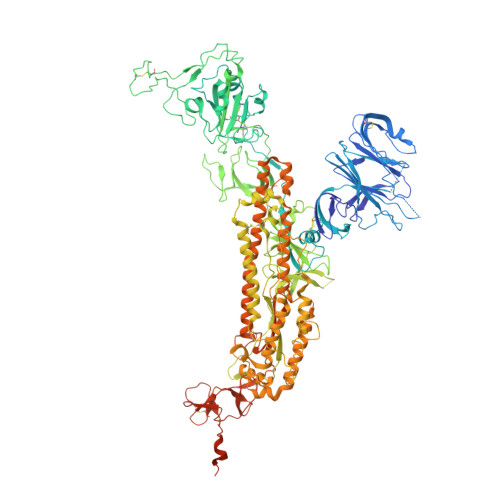
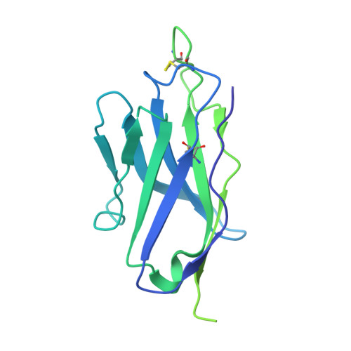
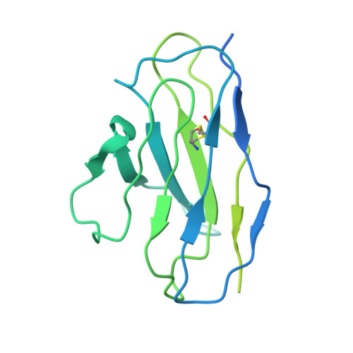
SARS-CoV-2 rapidly mutates and acquires resistance to neutralizing antibodies. We report an in-silico-designed antibody that restores the neutralizing activity of a neutralizing antibody. Our previously generated antibody, UT28K, exhibited broad neutralizing activity against mutant variants; however, its efficacy against Omicron BA.1 was compromised by the mutation. Using previously determined structural information, we designed a modified-UT28K (V H T28R/N57D), UT28K-RD targeting the mutation site. In vitro and in vivo experiments demonstrated the efficacy of UT28K-RD in neutralizing Omicron BA.1. Although the experimentally determined structure partially differed from the predicted model, our study serves as a successful case of antibody design, wherein the predicted amino acid substitution enhanced the recognition of the previously elusive Omicron BA.1. We anticipate that numerous similar cases will be reported, showcasing the potential of this approach for improving protein-protein interactions. Our findings will contribute to the development of novel therapeutic strategies for highly mutable viruses, such as SARS-CoV-2.
Organizational Affiliation:
Department of Immunology, Faculty of Medicine, Academic Assembly, University of Toyama, Toyama, Japan; Center for Advanced Antibody Drug Development, University of Toyama, Toyama, Japan. Electronic address: toz@med.u-toyama.ac.jp.
Institute for Integrated Cell-Material Sciences, Kyoto University, Yoshidahonnmachi, Sakyo-ku, Kyoto, Japan. Electronic address: ikeda.yoshiki.3r@kyoto-u.ac.jp.
Laboratory of Biomolecular Science, Faculty of Pharmaceutical Sciences, Hokkaido University, Sapporo, Japan.
Department of Microbiology and Immunology, Faculty of Medicine, Hokkaido University, Sapporo, Japan; Institute for Vaccine Research and Development (HU-IVReD), Hokkaido University, Sapporo, Japan.
Department of Cardiovascular Medicine, Graduate School of Medical Science, Kyoto Prefectural University of Medicine, Kyoto, Japan.
Department of Diagnostic Pathology, Faculty of Medicine, Academic Assembly, University of Toyama, Toyama, Japan.
Department of Virology, Toyama Institute of Health, Toyama, Japan.
Laboratory of Medical Virology, Institute for Life and Medical Sciences, Kyoto University, Kyoto, Japan.
Department of Nephrology, Graduate School of Medical Science, Kyoto Prefectural University of Medicine, Kyoto, Japan.
Laboratory of Biomolecular Science, Faculty of Pharmaceutical Sciences, Hokkaido University, Sapporo, Japan; Institute for Vaccine Research and Development (HU-IVReD), Hokkaido University, Sapporo, Japan; Center for Research and Education on Drug Discovery, Faculty of Pharmaceutical Sciences, Hokkaido University, Sapporo, Japan; Division of Pathogen Structure, International Institute for Zoonosis Control, Hokkaido University, Sapporo, Japan.
Laboratory of Biomolecular Science, Faculty of Pharmaceutical Sciences, Hokkaido University, Sapporo, Japan; Institute for Vaccine Research and Development (HU-IVReD), Hokkaido University, Sapporo, Japan; Center for Research and Education on Drug Discovery, Faculty of Pharmaceutical Sciences, Hokkaido University, Sapporo, Japan; Division of Pathogen Structure, International Institute for Zoonosis Control, Hokkaido University, Sapporo, Japan; Global Station for Biosurfaces and Drug Discovery, Hokkaido University, Sapporo, Japan.
Department of Microbiology and Immunology, Faculty of Medicine, Hokkaido University, Sapporo, Japan; Institute for Vaccine Research and Development (HU-IVReD), Hokkaido University, Sapporo, Japan; Laboratory of Virus Control, Research Institute for Microbial Diseases, Osaka University, Suita, Japan; AMED-CREST, Japan Agency for Medical Research and Development (AMED), Tokyo, Japan.
Department of Immunology, Faculty of Medicine, Academic Assembly, University of Toyama, Toyama, Japan; Center for Advanced Antibody Drug Development, University of Toyama, Toyama, Japan.
Center for Advanced Antibody Drug Development, University of Toyama, Toyama, Japan; Department of Clinical Laboratory and Molecular Pathology, Faculty of Medicine, Academic Assembly, University of Toyama, Toyama, Japan.
AA [auth A] BA [auth A] CA [auth A] DA [auth A] EA [auth A]
AA [auth A], BA [auth A], CA [auth A], DA [auth A], EA [auth A], FA [auth A], GA [auth A], HA [auth A], IA [auth A], JA [auth A], KA [auth A], LA [auth C], MA [auth C], N [auth B], NA [auth C], O [auth B], OA [auth C], P [auth B], PA [auth C], Q [auth B], QA [auth C], R [auth B], RA [auth C], S [auth B], SA [auth C], T [auth B], TA [auth C], U [auth B], UA [auth C], V [auth B], W [auth B], X [auth B], Y [auth B], Z [auth A]
2-acetamido-2-deoxy-beta-D-glucopyranose C8 H15 N O6 OVRNDRQMDRJTHS-FMDGEEDCSA-N