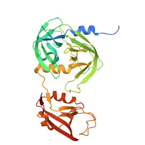
The structure of MucD from Pseudomonas syringae revealed N-terminal loop-mediated trimerization of HtrA-like serine protease.
Kim, J.H., Lee, G.H., Jeong, J.H., Kim, Y.G., Park, H.H.(2023) Biochem Biophys Res Commun 688: 149175-149175
- PubMed: 37976815 Search on PubMed
- DOI: https://doi.org/10.1016/j.bbrc.2023.149175
- Primary Citation Related Structures:
8K2Y - PubMed Abstract:
Protein quality control mechanisms are essential for maintaining cellular integrity, and the HtrA family of serine proteases plays a crucial role in handling folding stress in prokaryotic periplasm. Escherichia coli harbors three HtrA members, namely, DegS, DegP, and DegQ, which share a common domain structure. MucD, a putative HtrA family member that resembles DegP, is involved in alginate biosynthesis regulation and the stress response. Pseudomonas syringae causes plant diseases and opportunistic infections in humans. This study presents the high-resolution structure of MucD from Pseudomonas syringae (psMucD), revealing its composition as a typical HtrA family serine protease with protease and PDZ domains. Its findings suggest that psMucD containing one PDZ domain is a trimer in solution, and psMucD trimerization is mediated by its N-terminal loop. Sequence and structural analyses revealed similarities and differences with other HtrA family members. Additionally, this study provides a model of psMucD's catalytic process, comparing it with other members of the HtrA family of serine proteases.
- College of Pharmacy, Chung-Ang University, Seoul, 06974, Republic of Korea; Department of Global Innovative Drugs, Graduate School of Chung-Ang University, Seoul, 06974, Republic of Korea.
Organizational Affiliation: