


Crystal Structure of Rv0047c from Mycobacterium tuberculosis
Ansari, M.S., Yadav, V., Zohib, M., Pal, R.K., Biswal, B.K., Arora, A.To be published.
Experimental Data Snapshot
Starting Model: in silico
View more details
wwPDB Validation 3D Report Full Report

Entity ID: 1 | |||||
|---|---|---|---|---|---|
| Molecule | Chains | Sequence Length | Organism | Details | Image |
| Conserved protein | 182 | Mycobacterium tuberculosis H37Rv | Mutation(s): 0 Gene Names: Rv0047c | 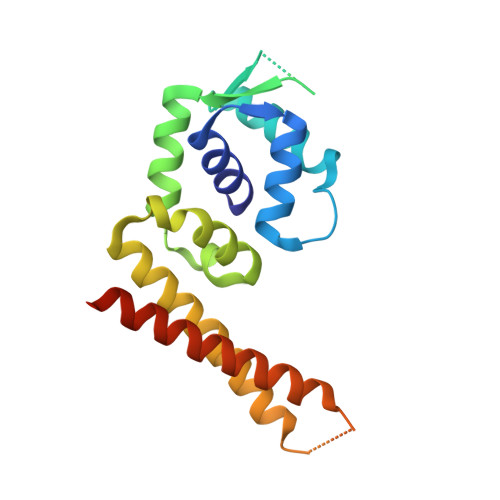 | |
UniProt | |||||
Entity Groups | |||||
| Sequence Clusters | 30% Identity50% Identity70% Identity90% Identity95% Identity100% Identity | ||||
| UniProt Group | P71704 | ||||
Sequence AnnotationsExpand | |||||
Reference Sequence | |||||
| Length ( Å ) | Angle ( ˚ ) |
|---|---|
| a = 102.17 | α = 90 |
| b = 102.17 | β = 90 |
| c = 191.88 | γ = 120 |
| Software Name | Purpose |
|---|---|
| REFMAC | refinement |
| Aimless | data scaling |
| iMOSFLM | data reduction |
| PHASER | phasing |
| Funding Organization | Location | Grant Number |
|---|---|---|
| Science and Engineering Research Board (SERB) | India | -- |


