8JOT

Crystal structure of CSF-1R kinase domain with sulfatinib


Experimental Data Snapshot

  • Method: X-RAY DIFFRACTION
  • Resolution: 1.69 Å
  • R-Value Free: 0.207 
  • R-Value Work: 0.179 
  • R-Value Observed: 0.180 

wwPDB Validation   3D Report Full Report


Ligand Structure Quality Assessment 


This is version 1.0 of the entry. See complete history


Literature

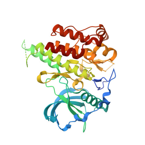
Structural basis and selectivity of sulfatinib binding to FGFR and CSF-1R.

Lin, Q.Dai, S.Qu, L.Lin, H.Guo, M.Wei, H.Chen, Y.Chen, X.

(2024) Commun Chem 7: 3-3

  • DOI: https://doi.org/10.1038/s42004-023-01084-0
  • Primary Citation of Related Structures:  
    8JMZ, 8JOT

  • PubMed Abstract: 

    Acquired drug resistance poses a challenge for single-target FGFR inhibitors, leading to the development of dual- or multi-target FGFR inhibitors. Sulfatinib is a multi-target kinase inhibitor for treating neuroendocrine tumors, selectively targeting FGFR1/CSF-1R. To elucidate the molecular mechanisms behind its binding and kinase selectivity, we determined the crystal structures of sulfatinib with FGFR1/CSF-1R. The results reveal common structural features and distinct conformational adaptability of sulfatinib in response to FGFR1/CSF-1R binding. Further biochemical and structural analyses disclose sensitivity of sulfatinib to FGFR/CSF-1R gatekeeper mutations. The insensitivity of sulfatinib to FGFR gatekeeper mutations highlights the indispensable interactions with the hydrophobic pocket for FGFR selectivity, whereas the rotatory flexibility may enable sulfatinib to overcome CSF-1R T663I . This study not only sheds light on the structural basis governing sulfatinib's FGFR/CSF-1R inhibition, but also provides valuable insights into the rational design of dual- or multi-target FGFR inhibitors with selectivity for CSF-1R and sensitivity to gatekeeper mutations.


  • Organizational Affiliation

    Department of Oncology, NHC Key Laboratory of Cancer Proteomics, State Local Joint Engineering Laboratory for Anticancer Drugs, Xiangya Hospital, Central South University, Changsha, Hunan, 410008, China.


Macromolecules
Find similar proteins by:  (by identity cutoff)  |  3D Structure
Entity ID: 1
MoleculeChains Sequence LengthOrganismDetailsImage
Macrophage colony-stimulating factor 1 receptor354Homo sapiensMutation(s): 3 
Gene Names: CSF1RFMS
EC: 2.7.10.1
UniProt & NIH Common Fund Data Resources
Find proteins for P07333 (Homo sapiens)
Explore P07333 
Go to UniProtKB:  P07333
PHAROS:  P07333
GTEx:  ENSG00000182578 
Entity Groups  
Sequence Clusters30% Identity50% Identity70% Identity90% Identity95% Identity100% Identity
UniProt GroupP07333
Sequence Annotations
Expand
  • Reference Sequence
Small Molecules
Ligands 2 Unique
IDChains Name / Formula / InChI Key2D Diagram3D Interactions
UKI (Subject of Investigation/LOI)
Query on UKI

Download Ideal Coordinates CCD File 
B [auth A]N-(2-(dimethylamino)ethyl)-1-(3-((4-((2-methyl-1H-indol-5-yl)oxy)pyrimidin-2-yl)amino)phenyl)methanesulfonamide
C24 H28 N6 O3 S
TTZSNFLLYPYKIL-UHFFFAOYSA-N
GOL
Query on GOL

Download Ideal Coordinates CCD File 
C [auth A]GLYCEROL
C3 H8 O3
PEDCQBHIVMGVHV-UHFFFAOYSA-N
Experimental Data & Validation

Experimental Data

  • Method: X-RAY DIFFRACTION
  • Resolution: 1.69 Å
  • R-Value Free: 0.207 
  • R-Value Work: 0.179 
  • R-Value Observed: 0.180 
  • Space Group: P 43 21 2
Unit Cell:
Length ( Å )Angle ( ˚ )
a = 64.31α = 90
b = 64.31β = 90
c = 184.685γ = 90
Software Package:
Software NamePurpose
PHENIXrefinement
HKL-2000data reduction
HKL-2000data scaling
PHENIXphasing
PDB_EXTRACTdata extraction

Structure Validation

View Full Validation Report


Ligand Structure Quality Assessment 


Entry History & Funding Information

Deposition Data


Funding OrganizationLocationGrant Number
National Natural Science Foundation of China (NSFC)China82202920

Revision History  (Full details and data files)

  • Version 1.0: 2024-03-27
    Type: Initial release