8JNS

cryo-EM structure of a CED-4 hexamer


Experimental Data Snapshot

  • Method: ELECTRON MICROSCOPY
  • Resolution: 4.20 Å
  • Aggregation State: PARTICLE 
  • Reconstruction Method: SINGLE PARTICLE 

wwPDB Validation   3D Report Full Report


This is version 1.1 of the entry. See complete history


Literature

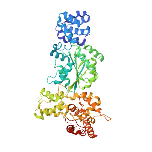
Structural insights into CED-3 activation.

Li, Y.Tian, L.Zhang, Y.Shi, Y.

(2023) Life Sci Alliance 6

  • DOI: https://doi.org/10.26508/lsa.202302056
  • Primary Citation of Related Structures:  
    8JNS, 8JO0, 8JOL

  • PubMed Abstract: 

    In Caenorhabditis elegans (C. elegans) , onset of programmed cell death is marked with the activation of CED-3, a process that requires assembly of the CED-4 apoptosome. Activated CED-3 forms a holoenzyme with the CED-4 apoptosome to cleave a wide range of substrates, leading to irreversible cell death. Despite decades of investigations, the underlying mechanism of CED-4-facilitated CED-3 activation remains elusive. Here, we report cryo-EM structures of the CED-4 apoptosome and three distinct CED-4/CED-3 complexes that mimic different activation stages for CED-3. In addition to the previously reported octamer in crystal structures, CED-4, alone or in complex with CED-3, exists in multiple oligomeric states. Supported by biochemical analyses, we show that the conserved CARD-CARD interaction promotes CED-3 activation, and initiation of programmed cell death is regulated by the dynamic organization of the CED-4 apoptosome.


  • Organizational Affiliation

    Beijing Frontier Research Center for Biological Structures, Tsinghua-Peking Center for Life Sciences, School of Life Sciences, Tsinghua University, Beijing, China liyini@mail.tsinghua.edu.cn.


Macromolecules
Find similar proteins by:  (by identity cutoff)  |  3D Structure
Entity ID: 1
MoleculeChains Sequence LengthOrganismDetailsImage
Cell death protein 4
A, B, C, D, E
A, B, C, D, E, F, G, H, I
549Caenorhabditis elegansMutation(s): 0 
Gene Names: ced-4C35D10.9
UniProt
Find proteins for P30429 (Caenorhabditis elegans)
Explore P30429 
Go to UniProtKB:  P30429
Entity Groups  
Sequence Clusters30% Identity50% Identity70% Identity90% Identity95% Identity100% Identity
UniProt GroupP30429
Sequence Annotations
Expand
  • Reference Sequence
Small Molecules
Ligands 2 Unique
IDChains Name / Formula / InChI Key2D Diagram3D Interactions
ATP (Subject of Investigation/LOI)
Query on ATP

Download Ideal Coordinates CCD File 
K [auth A]
M [auth B]
O [auth C]
Q [auth D]
S [auth E]
K [auth A],
M [auth B],
O [auth C],
Q [auth D],
S [auth E],
U [auth F]
ADENOSINE-5'-TRIPHOSPHATE
C10 H16 N5 O13 P3
ZKHQWZAMYRWXGA-KQYNXXCUSA-N
MG (Subject of Investigation/LOI)
Query on MG

Download Ideal Coordinates CCD File 
J [auth A]
L [auth B]
N [auth C]
P [auth D]
R [auth E]
J [auth A],
L [auth B],
N [auth C],
P [auth D],
R [auth E],
T [auth F]
MAGNESIUM ION
Mg
JLVVSXFLKOJNIY-UHFFFAOYSA-N
Experimental Data & Validation

Experimental Data

  • Method: ELECTRON MICROSCOPY
  • Resolution: 4.20 Å
  • Aggregation State: PARTICLE 
  • Reconstruction Method: SINGLE PARTICLE 

Structure Validation

View Full Validation Report


Entry History & Funding Information

Deposition Data

  • Released Date: 2023-06-28 
  • Deposition Author(s): Li, Y., Shi, Y.

Funding OrganizationLocationGrant Number
Other privateChina--

Revision History  (Full details and data files)

  • Version 1.0: 2023-06-28
    Type: Initial release
  • Version 1.1: 2023-08-02
    Changes: Database references