8JFK

PhK holoenzyme in inactive state, muscle isoform


Experimental Data Snapshot

  • Method: ELECTRON MICROSCOPY
  • Resolution: 2.90 Å
  • Aggregation State: PARTICLE 
  • Reconstruction Method: SINGLE PARTICLE 

wwPDB Validation   3D Report Full Report


This is version 1.1 of the entry. See complete history


Literature

Architecture and activation of human muscle phosphorylase kinase.

Yang, X.Zhu, M.Lu, X.Wang, Y.Xiao, J.

(2024) Nat Commun 15: 2719-2719

  • DOI: https://doi.org/10.1038/s41467-024-47049-2
  • Primary Citation of Related Structures:  
    8JFK, 8JFL, 8XY7, 8XYA, 8XYB

  • PubMed Abstract: 

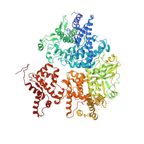
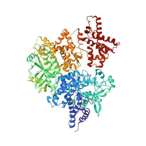
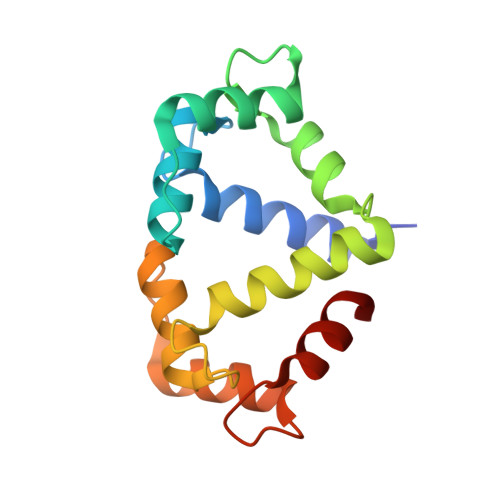
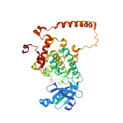
    The study of phosphorylase kinase (PhK)-regulated glycogen metabolism has contributed to the fundamental understanding of protein phosphorylation; however, the molecular mechanism of PhK remains poorly understood. Here we present the high-resolution cryo-electron microscopy structures of human muscle PhK. The 1.3-megadalton PhK α 4 β 4 γ 4 δ 4 hexadecamer consists of a tetramer of tetramer, wherein four αβγδ modules are connected by the central β 4 scaffold. The α- and β-subunits possess glucoamylase-like domains, but exhibit no detectable enzyme activities. The α-subunit serves as a bridge between the β-subunit and the γδ subcomplex, and facilitates the γ-subunit to adopt an autoinhibited state. Ca 2+ -free calmodulin (δ-subunit) binds to the γ-subunit in a compact conformation. Upon binding of Ca 2+ , a conformational change occurs, allowing for the de-inhibition of the γ-subunit through a spring-loaded mechanism. We also reveal an ADP-binding pocket in the β-subunit, which plays a role in allosterically enhancing PhK activity. These results provide molecular insights of this important kinase complex.


  • Organizational Affiliation

    State Key Laboratory of Protein and Plant Gene Research, School of Life Sciences, Peking University, Beijing, P.R. China.


Macromolecules
Find similar proteins by:  (by identity cutoff)  |  3D Structure
Entity ID: 1
MoleculeChains Sequence LengthOrganismDetailsImage
Phosphorylase b kinase regulatory subunit betaA [auth B],
D [auth N],
E [auth J],
G [auth F]
1,093Homo sapiensMutation(s): 0 
Gene Names: PHKB
UniProt & NIH Common Fund Data Resources
Find proteins for Q93100 (Homo sapiens)
Explore Q93100 
Go to UniProtKB:  Q93100
PHAROS:  Q93100
GTEx:  ENSG00000102893 
Entity Groups  
Sequence Clusters30% Identity50% Identity70% Identity90% Identity95% Identity100% Identity
UniProt GroupQ93100
Sequence Annotations
Expand
  • Reference Sequence
Find similar proteins by:  (by identity cutoff)  |  3D Structure
Entity ID: 2
MoleculeChains Sequence LengthOrganismDetailsImage
Phosphorylase b kinase regulatory subunit alpha, skeletal muscle isoformB [auth M],
C [auth A],
F [auth I],
H [auth E]
1,223Homo sapiensMutation(s): 0 
Gene Names: PHKA1PHKA
UniProt & NIH Common Fund Data Resources
Find proteins for P46020 (Homo sapiens)
Explore P46020 
Go to UniProtKB:  P46020
PHAROS:  P46020
GTEx:  ENSG00000067177 
Entity Groups  
Sequence Clusters30% Identity50% Identity70% Identity90% Identity95% Identity100% Identity
UniProt GroupP46020
Sequence Annotations
Expand
  • Reference Sequence
Find similar proteins by:  (by identity cutoff)  |  3D Structure
Entity ID: 3
MoleculeChains Sequence LengthOrganismDetailsImage
Calmodulin-1I [auth D],
K [auth H],
M [auth L],
O [auth P]
149Homo sapiensMutation(s): 0 
Gene Names: CALM1CALMCAMCAM1
UniProt & NIH Common Fund Data Resources
Find proteins for P0DP23 (Homo sapiens)
Explore P0DP23 
Go to UniProtKB:  P0DP23
PHAROS:  P0DP23
GTEx:  ENSG00000198668 
Entity Groups  
Sequence Clusters30% Identity50% Identity70% Identity90% Identity95% Identity100% Identity
UniProt GroupP0DP23
Sequence Annotations
Expand
  • Reference Sequence
Find similar proteins by:  (by identity cutoff)  |  3D Structure
Entity ID: 4
MoleculeChains Sequence LengthOrganismDetailsImage
Phosphorylase b kinase gamma catalytic chain, skeletal muscle/heart isoformJ [auth C],
L [auth G],
N [auth K],
P [auth O]
387Homo sapiensMutation(s): 0 
Gene Names: PHKG1PHKG
EC: 2.7.11.19 (PDB Primary Data), 2.7.11.1 (PDB Primary Data), 2.7.11.26 (PDB Primary Data)
UniProt & NIH Common Fund Data Resources
Find proteins for Q16816 (Homo sapiens)
Explore Q16816 
Go to UniProtKB:  Q16816
PHAROS:  Q16816
GTEx:  ENSG00000164776 
Entity Groups  
Sequence Clusters30% Identity50% Identity70% Identity90% Identity95% Identity100% Identity
UniProt GroupQ16816
Sequence Annotations
Expand
  • Reference Sequence
Experimental Data & Validation

Experimental Data

  • Method: ELECTRON MICROSCOPY
  • Resolution: 2.90 Å
  • Aggregation State: PARTICLE 
  • Reconstruction Method: SINGLE PARTICLE 
EM Software:
TaskSoftware PackageVersion
MODEL REFINEMENTPHENIX1.19.2_4158:

Structure Validation

View Full Validation Report


Entry History & Funding Information

Deposition Data


Funding OrganizationLocationGrant Number
National Natural Science Foundation of China (NSFC)China--

Revision History  (Full details and data files)

  • Version 1.0: 2024-04-03
    Type: Initial release
  • Version 1.1: 2024-04-10
    Changes: Database references