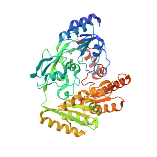
Lactate dehydrogenase D is a general dehydrogenase for D-2-hydroxyacids and is associated with D-lactic acidosis.
Jin, S., Chen, X., Yang, J., Ding, J.(2023) Nat Commun 14: 6638-6638
- PubMed: 37863926 Search on PubMedSearch on PubMed Central
- DOI: https://doi.org/10.1038/s41467-023-42456-3
- Primary Citation Related Structures:
8JDB, 8JDC, 8JDD, 8JDE, 8JDF, 8JDG, 8JDN, 8JDO, 8JDP, 8JDQ, 8JDR, 8JDS, 8JDT, 8JDU, 8JDV, 8JDX, 8JDY, 8JDZ - PubMed Abstract:
Mammalian lactate dehydrogenase D (LDHD) catalyzes the oxidation of D-lactate to pyruvate. LDHD mutations identified in patients with D-lactic acidosis lead to deficient LDHD activity. Here, we perform a systematic biochemical study of mouse LDHD (mLDHD) and determine the crystal structures of mLDHD in FAD-bound form and in complexes with FAD, Mn 2+ and a series of substrates or products. We demonstrate that mLDHD is an Mn 2+ -dependent general dehydrogenase which exhibits catalytic activity for D-lactate and other D-2-hydroxyacids containing hydrophobic moieties, but no activity for their L-isomers or D-2-hydroxyacids containing hydrophilic moieties. The substrate-binding site contains a positively charged pocket to bind the common glycolate moiety and a hydrophobic pocket with some elasticity to bind the varied hydrophobic moieties of substrates. The structural and biochemical data together reveal the molecular basis for the substrate specificity and catalytic mechanism of LDHD, and the functional roles of mutations in the pathogenesis of D-lactic acidosis.
- State Key Laboratory of Molecular Biology, Shanghai Institute of Biochemistry and Cell Biology, Center for Excellence in Molecular Cell Science, University of Chinese Academy of Sciences, Chinese Academy of Sciences, 320 Yue-Yang Road, Shanghai, 200031, China.
Organizational Affiliation: