8JD5

Cryo-EM structure of Gi1-bound mGlu2-mGlu4 heterodimer


Experimental Data Snapshot

  • Method: ELECTRON MICROSCOPY
  • Resolution: 3.60 Å
  • Aggregation State: PARTICLE 
  • Reconstruction Method: SINGLE PARTICLE 

wwPDB Validation   3D Report Full Report


This is version 1.1 of the entry. See complete history


Literature

Structural insights into dimerization and activation of the mGlu2-mGlu3 and mGlu2-mGlu4 heterodimers.

Wang, X.Wang, M.Xu, T.Feng, Y.Shao, Q.Han, S.Chu, X.Xu, Y.Lin, S.Zhao, Q.Wu, B.

(2023) Cell Res 33: 762-774

  • DOI: https://doi.org/10.1038/s41422-023-00830-2
  • Primary Citation of Related Structures:  
    8JCU, 8JCV, 8JCW, 8JCX, 8JCY, 8JCZ, 8JD0, 8JD1, 8JD2, 8JD3, 8JD4, 8JD5, 8JD6

  • PubMed Abstract: 

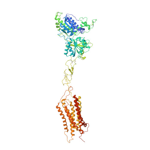
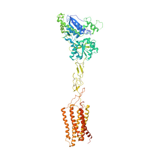
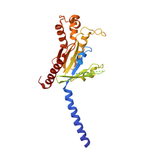
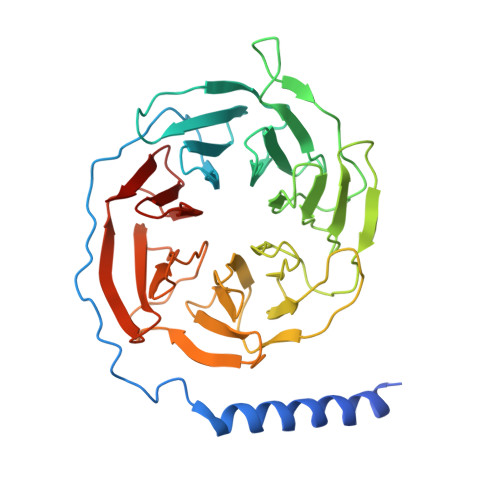
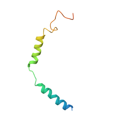
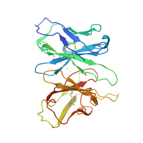
    Heterodimerization of the metabotropic glutamate receptors (mGlus) has shown importance in the functional modulation of the receptors and offers potential drug targets for treating central nervous system diseases. However, due to a lack of molecular details of the mGlu heterodimers, understanding of the mechanisms underlying mGlu heterodimerization and activation is limited. Here we report twelve cryo-electron microscopy (cryo-EM) structures of the mGlu2-mGlu3 and mGlu2-mGlu4 heterodimers in different conformational states, including inactive, intermediate inactive, intermediate active and fully active conformations. These structures provide a full picture of conformational rearrangement of mGlu2-mGlu3 upon activation. The Venus flytrap domains undergo a sequential conformational change, while the transmembrane domains exhibit a substantial rearrangement from an inactive, symmetric dimer with diverse dimerization patterns to an active, asymmetric dimer in a conserved dimerization mode. Combined with functional data, these structures reveal that stability of the inactive conformations of the subunits and the subunit-G protein interaction pattern are determinants of asymmetric signal transduction of the heterodimers. Furthermore, a novel binding site for two mGlu4 positive allosteric modulators was observed in the asymmetric dimer interfaces of the mGlu2-mGlu4 heterodimer and mGlu4 homodimer, and may serve as a drug recognition site. These findings greatly extend our knowledge about signal transduction of the mGlus.


  • Organizational Affiliation

    State Key Laboratory of Drug Research, Shanghai Institute of Materia Medica, Chinese Academy of Sciences, Shanghai, China.


Macromolecules
Find similar proteins by:  (by identity cutoff)  |  3D Structure
Entity ID: 1
MoleculeChains Sequence LengthOrganismDetailsImage
Metabotropic glutamate receptor 2A [auth 2]870Homo sapiensMutation(s): 0 
Gene Names: GRM2GPRC1BMGLUR2
Membrane Entity: Yes 
UniProt & NIH Common Fund Data Resources
Find proteins for Q14416 (Homo sapiens)
Explore Q14416 
Go to UniProtKB:  Q14416
PHAROS:  Q14416
GTEx:  ENSG00000164082 
Entity Groups  
Sequence Clusters30% Identity50% Identity70% Identity90% Identity95% Identity100% Identity
UniProt GroupQ14416
Sequence Annotations
Expand
  • Reference Sequence
Find similar proteins by:  (by identity cutoff)  |  3D Structure
Entity ID: 2
MoleculeChains Sequence LengthOrganismDetailsImage
Metabotropic glutamate receptor 4B [auth 4]916Homo sapiensMutation(s): 0 
Gene Names: GRM4GPRC1DMGLUR4
Membrane Entity: Yes 
UniProt & NIH Common Fund Data Resources
Find proteins for Q14833 (Homo sapiens)
Explore Q14833 
Go to UniProtKB:  Q14833
PHAROS:  Q14833
GTEx:  ENSG00000124493 
Entity Groups  
Sequence Clusters30% Identity50% Identity70% Identity90% Identity95% Identity100% Identity
UniProt GroupQ14833
Sequence Annotations
Expand
  • Reference Sequence
Find similar proteins by:  (by identity cutoff)  |  3D Structure
Entity ID: 3
MoleculeChains Sequence LengthOrganismDetailsImage
Guanine nucleotide-binding protein G(i) subunit alpha-1C [auth A]354Homo sapiensMutation(s): 4 
Gene Names: GNAI1
Membrane Entity: Yes 
UniProt & NIH Common Fund Data Resources
Find proteins for P63096 (Homo sapiens)
Explore P63096 
Go to UniProtKB:  P63096
PHAROS:  P63096
GTEx:  ENSG00000127955 
Entity Groups  
Sequence Clusters30% Identity50% Identity70% Identity90% Identity95% Identity100% Identity
UniProt GroupP63096
Sequence Annotations
Expand
  • Reference Sequence
Find similar proteins by:  (by identity cutoff)  |  3D Structure
Entity ID: 4
MoleculeChains Sequence LengthOrganismDetailsImage
Guanine nucleotide-binding protein G(I)/G(S)/G(T) subunit beta-1D [auth B]351Homo sapiensMutation(s): 0 
Gene Names: GNB1
UniProt & NIH Common Fund Data Resources
Find proteins for P62873 (Homo sapiens)
Explore P62873 
Go to UniProtKB:  P62873
PHAROS:  P62873
GTEx:  ENSG00000078369 
Entity Groups  
Sequence Clusters30% Identity50% Identity70% Identity90% Identity95% Identity100% Identity
UniProt GroupP62873
Sequence Annotations
Expand
  • Reference Sequence
Find similar proteins by:  (by identity cutoff)  |  3D Structure
Entity ID: 5
MoleculeChains Sequence LengthOrganismDetailsImage
Guanine nucleotide-binding protein G(I)/G(S)/G(O) subunit gamma-2E [auth C]71Homo sapiensMutation(s): 0 
Gene Names: GNG2
Membrane Entity: Yes 
UniProt & NIH Common Fund Data Resources
Find proteins for P59768 (Homo sapiens)
Explore P59768 
Go to UniProtKB:  P59768
PHAROS:  P59768
GTEx:  ENSG00000186469 
Entity Groups  
Sequence Clusters30% Identity50% Identity70% Identity90% Identity95% Identity100% Identity
UniProt GroupP59768
Sequence Annotations
Expand
  • Reference Sequence
Find similar proteins by:  (by identity cutoff)  |  3D Structure
Entity ID: 6
MoleculeChains Sequence LengthOrganismDetailsImage
scFvF [auth H]257Escherichia coliMutation(s): 0 
Entity Groups  
Sequence Clusters30% Identity50% Identity70% Identity90% Identity95% Identity100% Identity
Sequence Annotations
Expand
  • Reference Sequence
Small Molecules
Ligands 6 Unique
IDChains Name / Formula / InChI Key2D Diagram3D Interactions
PEF
Query on PEF

Download Ideal Coordinates CCD File 
J [auth 2]DI-PALMITOYL-3-SN-PHOSPHATIDYLETHANOLAMINE
C37 H74 N O8 P
SLKDGVPOSSLUAI-PGUFJCEWSA-N
CLR
Query on CLR

Download Ideal Coordinates CCD File 
I [auth 2],
L [auth 2],
M [auth 2],
N [auth 4],
R [auth 4]
CHOLESTEROL
C27 H46 O
HVYWMOMLDIMFJA-DPAQBDIFSA-N
HZR (Subject of Investigation/LOI)
Query on HZR

Download Ideal Coordinates CCD File 
G [auth 2]1-butyl-3-chloranyl-4-(4-phenylpiperidin-1-yl)pyridin-2-one
C20 H25 Cl N2 O
HYOGJHCDLQSAHX-UHFFFAOYSA-N
BQI (Subject of Investigation/LOI)
Query on BQI

Download Ideal Coordinates CCD File 
Q [auth 4]5-methyl-N-(4-methylpyrimidin-2-yl)-4-(1H-pyrazol-4-yl)-1,3-thiazol-2-amine
C12 H12 N6 S
MIQNXKWDQRNHAU-UHFFFAOYSA-N
NAG
Query on NAG

Download Ideal Coordinates CCD File 
K [auth 2],
P [auth 4]
2-acetamido-2-deoxy-beta-D-glucopyranose
C8 H15 N O6
OVRNDRQMDRJTHS-FMDGEEDCSA-N
GLU (Subject of Investigation/LOI)
Query on GLU

Download Ideal Coordinates CCD File 
H [auth 2],
O [auth 4]
GLUTAMIC ACID
C5 H9 N O4
WHUUTDBJXJRKMK-VKHMYHEASA-N
Experimental Data & Validation

Experimental Data

  • Method: ELECTRON MICROSCOPY
  • Resolution: 3.60 Å
  • Aggregation State: PARTICLE 
  • Reconstruction Method: SINGLE PARTICLE 

Structure Validation

View Full Validation Report


Entry History & Funding Information

Deposition Data


Funding OrganizationLocationGrant Number
National Science Foundation (NSF, China)China31825010
National Science Foundation (NSF, China)China82121005

Revision History  (Full details and data files)

  • Version 1.0: 2023-06-21
    Type: Initial release
  • Version 1.1: 2023-10-18
    Changes: Data collection, Database references