Structural basis for the development of potential inhibitors targeting FadD23 from Mycobacterium tuberculosis.
Yan, M., Ma, M., Chen, R., Cao, Y., Zhang, W., Liu, X.(2023) Acta Crystallogr F Struct Biol Commun 79: 208-216
- PubMed: 37522751 Search on PubMedSearch on PubMed Central
- DOI: https://doi.org/10.1107/S2053230X23005836
- Primary Citation Related Structures:
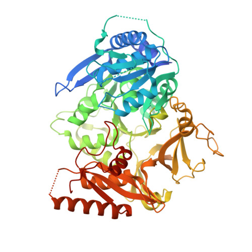
8IQU - PubMed Abstract:
Sulfolipid-1 (SL-1) is a lipid that is abundantly found in the cell wall of Mycobacterium tuberculosis (Mtb). MtbFadD23 is crucial in the SL-1 synthesis pathway. Previously, 5'-O-[N-(11-phenoxyundecanoyl)sulfamoyl]adenosine (PhU-AMS) has been shown to be a general inhibitor of fatty-acid-adenylating enzymes (FadDs) in Mtb. However, the fatty acyl-AMP ligase (FAAL) class of FadDs, which includes MtbFadD23, appears to be functionally nonredundant in the production of multiple fatty acids. In this study, the ability of PhU-AMS to bind to MtbFadD23 was examined under in vitro conditions. The crystal structure of the MtbFadD23-PhU-AMS complex was determined at a resolution of 2.64 Å. Novel features were identified by structural analysis and comparison. Although PhU-AMS could bind to MtbFadD23, it did not inhibit the FAAL adenylation activity of MtbFadD23. However, PhU-AMS improved the main T m value in a differential scanning fluorimetry assay, and a structural comparison of MtbFadD23-PhU-AMS with FadD32 and PA1221 suggested that PhU-AMS blocks the loading of the acyl chain onto Pks2. This study sheds light on the structure-based design of specific inhibitors of MtbFadD23 and general inhibitors of FAALs.
- State Key Laboratory of Medicinal Chemical Biology, Frontiers Science Center for Cell Responses, College of Life Sciences, Nankai University, Tianjin, People's Republic of China.
Organizational Affiliation: